Posts

28 November 2019

A study recently published in the New England Journal of Medicine* suggests that the use of breast MRI in addition to mammograms in women with extremely dense breast can improve the rate of “interval cancer” detection.

Interval cancers are those that occur after an apparently normal mammogram, but prior to the next scheduled mammogram. An example could be a lump felt by a patient a few months after a “clear” mammogram. Interval cancers are more likely to be more aggressive (triple negative or Her2/neu over-expressed) and/or occur in women who have dense breasts on mammogram.

Researchers in the Netherlands evaluated the role of breast MRI as a supplemental screening tool in women age 50-74, who had a normal mammogram and an “extremely dense” (BIRADS 4 density) reading. After a normal mammogram, women were randomized 1:4 to supplemental MRI versus continued mammogram only (1 woman received an invitation for MRI for every 4 who continued undergoing only mammograms). The standard mammography screening in the Netherlands is every 2 years. 8061 women were in the “intent to treat” with MRI group and 32,312 in the mammogram alone group. 59% (4783) of women randomized to the MRI group actually accepted and underwent supplemental MRI.

Some of the key findings:

  • In the MRI “intent to treat” group, they found 2.5 / 1000 interval cancers and in the mammography group there were 5.0 / 1000 interval cancers
  • However, out of the 20 interval cancers discovered in the MRI “intent to treat” group, only 4 were in women who had actually undergone MRI. The other 16 were in women were randomized to the MRI group but did not have the study – they had mammograms only
  • Out of the women who underwent MRI, approximately 10% (454) were recalled for additional imaging
  • 300 women had needle biopsies due to findings on MRI, and out of those, 79 (26%) were found to have cancer. 64 of the cancers were invasive and 15 were noninvasive (DCIS). 74% of the biopsies did NOT show cancer
  • The cancers detected by MRI on average were smaller than those detected by mammography. The majority were hormone receptor positive and lymph node negative
  • DCIS was more likely to be detected in women undergoing MRI (0.31%) versus mammogram alone (0.03%)
  • 0.1% of patients who had an MRI experienced an adverse reaction including fainting, contrast (dye) reaction, and issues with the intravenous line
  • The study was NOT designed to determine if MRI improved the survival rates from the cancers detected.

Some important points were raised in the accompanying editorial by Dr. Dan Longo*. He noted that with any screening test, the goal should be that early detection of the cancer results in a higher likelihood of cure but admitted that any screening test is associated with false positives, and many patients with an abnormal test will not have cancer. He also noted that an “imprecisely defined fraction of cancers that are detected by screening do not progress to cause symptomatic disease or to kill the patient” but that “our dilemma is that for most tumors we cannot tell the difference between cancers that can kill you and those that cannot.”

The study focused on patients with extremely dense breasts, which the authors estimate account for approximately 10% of patients undergoing mammograms. It is not clear if these findings would be similar in patients with level 3 breast density (heterogeneously dense), those undergoing tomosynthesis (3D) mammograms, or in patients who are undergoing mammograms every year. They did not stratify patients by risk assessment and it’s important to note that while dense breasts are a risk factor for breast cancer, not all women with dense breasts are considered “high risk”. US national guidelines currently recommend supplemental imaging with MRI in patients who have a lifetime risk of breast cancer of 20% or greater as calculated by a risk assessment model. Due to high recall and false positive rates associated with MRI as well as possible concerns related to the contrast material, we do not currently recommend MRI for all women, or even for all women with dense breasts.

As one of their concluding statements, the authors did note that the 10% recall rate “is a concern for potential implementation of supplemental screening” [with MRI]. This study adds to the body of literature regarding cancer detection in women with dense breasts, but Dr. Longo concluded in his editorial: “… will we be putting these women at increased risk of procedures without contributing to their eventual survival?” Going forward, it will be important to look at ways to decrease false positive results and costs of supplemental imaging, as well as evaluate whether or not outcomes are improved by additional testing.

*If you are not able to access the full study and would like a copy, please email me: contact at drattai dot com

5 July 2019

A study recently published in The Lancet Oncology found that the addition of MRI to mammography screening in high-risk patients detected breast cancers in earlier stages, but found that this technology was associated with a higher likelihood of false-positive results.

The study was performed at 12 hospitals in the Netherlands. Patients who were age 30-55 with a lifetime risk of breast cancer greater than 20% due to family history, but who tested negative for deleterious mutations in BRCA 1/2 and TP53 genes, were eligible to participate. Patients were randomized to one of the following surveillance arms:

  • Annual MRI, annual clinical breast exam, every other year mammogram [MRI-MG], or
  • Annual mammogram and annual clinical breast exam [MG]

1355 women were randomized and 231 were registered – the patients who were registered were those who did not provide consent for randomization, but followed one or the other protocol. Results from the patients who were registered but not randomized were used only in a portion of the data analysis.  

The authors found that over the 7-year study period:

  • 40 breast cancers were detected in the MRI-MG group, 15 were detected in the MG group
  • 24 invasive breast cancers were detected in the MRI-MG group, 8 were detected in the MG group
  • Invasive breast cancers detected in the MRI-MG group were smaller (9 versus 17 millimeters) than those in the MG group
  • Lymph node involvement was less likely to be seen in the MRI-MG group (4 of 24 cases, 17%) versus the MG group (5 of 8 cases, 63%)
  • There was one interval cancer (detected between screening exams) in the MRI-MG group and 2 in the MG group
  • Increased breast density was associated with higher stage at diagnosis in both study groups
  • Clinical breast exams generated a large number of false-positive results in both groups and detected only one cancer.

This study adds to the evidence nothing that the addition of MRI to screening mammography will increase the rate of cancer detection. National guidelines in the US do recommend breast MRI on an annual basis, in addition to annual mammography, for patients with a lifetime risk of 20% or greater. This study was not designed to assess outcomes for the patients who were diagnosed with breast cancer, but the authors postulated that MRI screening might lead to mortality reduction due to earlier stage at diagnosis. As reduction in mortality is the goal of screening, they stated that they plan to link the current study results with their national cancer database, and eventually publish 10-year mortality statistics for each of the groups. 

Additional Information: ASCO Post

18 September 2017

Breast MRIs are used in some newly diagnosed breast cancer patients, especially those with dense breast tissue. In addition, they are used as a supplement to mammography in women who are found to be at high risk. In order to distinguish between normal and cancerous tissue, an intravenous contrast dye is used. For MRI studies, the contrast material that is used is gadolinium-based.

Gadolinium is a heavy metal, and it has the potential to be retained in the brain and kidneys. Some recent studies in both the scientific and lay press have focused on the safety of gadolinium. While there have only been scattered reports of adverse events, an FDA advisory panel voted for warning labels and for more study.

The FDA advisory focuses primarily on a class of agents known as “linear”. The “macrocyclic” gadolinium contrast agents do not appear to have the same risk of depositing in the brain or kidneys. Both types of gadolinium contrast can be used for breast MRI studies. If you have concerns, ask the facility which form of gadolinium they use.

*Note – UCLA facilities (Westwood, Santa Monica and Santa Clarita), San Fernando Interventional Radiology (a RadNet facility) and Providence St. Joseph Hospital all use gadavist, a “macrocyclic” form of  gadolinium contrast, for their breast MRI studies. Because facility practice patterns may change over time, ask at the time of your exam if you have concerns.

24 September 2015

It is not uncommon that after a breast cancer diagnosis, a breast MRI is recommended. We know that there are limitations to mammography – it might not show everything of concern, especially in women with dense breast tissue. The idea behind preoperative MRI is that if there are other lesions, better to know about them and determine if they are cancerous or not, before going to surgery. This should lead to better surgical outcomes. Makes sense.

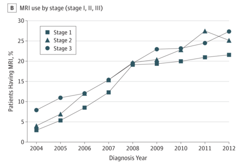
The problem is, studies have not been able to show a benefit to preoperative MRI. This was addressed in a recent issue of JAMA Oncology. Dr. Angel Arnaout and colleagues reported on the Use of Preoperative Magnetic Resonance Imaging for Breast Cancer: A Canadian Population-Based Study. In this retrospective population-based study, 53,015 patients with operable (early-stage) breast cancer were identified between 2003-2012.

Overall, close to 15% of patients underwent an MRI, and the use increased from 3% to 24% during the 10 year period.

Screen Shot 2015-10-15 at 2.08.59 PM

Patients undergoing a preoperative MRI were more likely to be younger, of higher socioeconomic status, cared for in teaching hospitals, being cared for surgeons with a high volume of breast cancer cases, and cared for by younger surgeons. Preoperative MRI was associated with a higher likelihood of additional imaging and biopsies, greater than 30 day wait time to surgery, and higher rates of mastectomy as well as contralateral prophylactic mastectomy.

These findings were discussed by Drs. Habib Rahbar and Constance Lehman in an accompanying editorial. They pointed out several limitations in the study, such as not knowing the MRI results, and not having the genetic mutations status (such as BRCA 1/2) of the patients. However, they noted that “this study adds to the growing body of evidence that the use of MRI in the preoperative setting is associated with more aggressive surgery of the affected breast”. They also noted that given concerns of breast cancer overtreatment, we need to modify our approach. They pointed out that MRI might be useful in developing individualized treatment approaches, such as multiple lumpectomies if more than one cancer is found (instead of mastectomy). They appropriately called for more research to determine how preoperative breast MRI should best be utilized.