26 February 2019

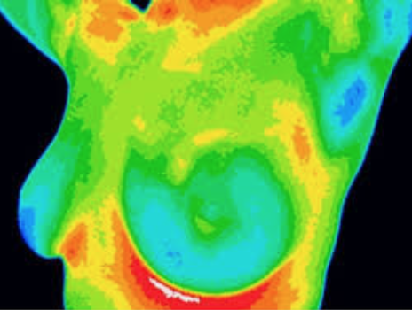
The US Food and Drug Administration (FDA) has issued a safety communication regarding the use of breast thermography as a sole means of breast cancer screening or diagnosis.

Thermography has been around for many years, and as seen by the FDA communication, has been the subject of multiple warning letters. Thermography works by detecting patters of heat flow on or just below the body surface. We know that breast (and other cancers) tend to have a more robust blood supply compared to normal tissue – and proponents of thermography feel that this reliably translates into detectable changes in heat patterns.

Thermography does not utilize radiation and there is no compression of the breast, making it appear to be an ideal alternative to mammography. Unfortunately, it has never been shown to reliably detect breast cancer, even in patients with suspicious mammography findings (see Collette, AE et al. paper referenced below).

The FDA alert notes that thermography was never approved as a stand alone test to screen for breast cancer, even though many facilities advertise it as an alternative to mammography and other imaging. Thermography practitioners also claim that precancerous changes can be detected many years prior to the development of cancer. I’ve seen many patients over the years who have had abnormal thermograms – and they leave the facility with recommendations including hormone creams (to apply to the breast), lymphatic massage, and repeat thermography in 3 months. Some of these patients in fact had large, advanced cancers. Thermography is not covered by insurance, and costs can range anywhere from $150 – 500 / session. While every thermogram report I’ve seen includes a recommendation for additional imaging with mammogram, ultrasound or MRI (usually in the fine print), this is never really discussed with the patient.

Mammograms have their flaws and early detection does not necessarily result in cure. However, thermography is not the answer.

Collette AE et al. 2014: Infrared Imaging does not Predict the Presence of Malignancy in Patients with Suspicious Radiologic Breast Abnormalities

GMA – Breast Cancer Survivor Shares Cautionary Tale

10 May 2018

The American Society of Breast Surgeons held their Annual Meeting in Orlando, FL from May 2nd – 6th. As usual, it was well attended – the meeting is known for being very practical and full of information that breast surgeons can bring back to their practices to help improve patient care.

I’ve picked a few topics to highlight in this post: Genetics, Imaging, Local Therapy, Systemic Therapy, Immunotherapy, Liquid Biopsy, Diet and Hormone Therapy, and Changing Paradigms. The following are comments expressed by the meeting speakers. My own comments will be noted in bold italics.

Genetics:

  • BRCA 1 mutation carriers are more likely to have triple negative breast cancer.
  • BRCA 2 mutation carriers are more likely to have ER positive, Her2/neu negative breast cancers.
  • The risk of a 2nd breast cancer in BRCA mutation carriers on average is about 2% per year depending on the specific mutation and the age of affected relatives. It can approach 60-80% in some patients. This increased risk of a new breast cancer is why bilateral mastectomy is often recommended. Removal of the opposite breast may result in improved overall survival but results from studies are mixed.
  • For BRCA mutation carriers, it is recommended that clinical breast exam (breast exam by the physician) be performed every 6-12 months. From age 25-29 annual MRI is recommended, and from age 30-75 annual mammogram (3D mammogram or tomosynthesis was recommended) along with MRI was recommended. It was stated that this screening regimen has not been shown to improve survival, but the screen-detected cancers were less likely to have lymph node involvement. No specific recommendation was made for imaging or exam after bilateral mastectomy.
  • MRI every 6 months has been suggested by some, but there are concerns about gadolinium (a heavy metal material which is the contrast agent used for breast MRI) buildup.
  • Removal of the ovaries is recommended around age 40.
  • In patients with BRCA mutations who undergo salpingo-oophorectomy (removal of the ovaries and fallopian tubes), estrogen replacement therapy has not been shown to increase subsequent breast cancer risk. However, combined estrogen / progesterone therapy may increase subsequent breast cancer risk. It was suggested to consider removing the uterus at the time of ovary removal, so that estrogen alone could be used (if the uterus is not removed, estrogen alone could increase the risk of uterine cancer).
  • There are many other genetic mutations that have been identified that have a variable association with increased breast cancer risk. It was stressed that family history and other factors need to be considered when these less common mutations (such as CHEK2, ATM, PALB2 and many more) are present, before recommending mastectomy.
  • It was stressed that the presence of a variant of unknown significance (VUS) should NOT prompt aggressive surgery.
  • A study was presented that demonstrated that current breast cancer genetic testing guidelines exclude almost half of high-risk patients, and a recommendation was made for testing of all breast cancer patients regardless of age, family history or other factors.

Breast Imaging:

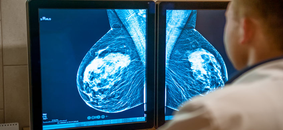
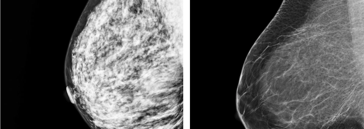
  • Dense breast (as determined by mammogram) reduces the sensitivity of mammograms, and also is associated with an increased risk of breast cancer.
  • It was stressed that determination of breast density is subjective and studies have shown significant variability in grading of breast density. Automated methods of assessing density are being evaluated.
  • 34 states have dense breast notification legislation. Some have supplemental screening (such as ultrasound) legislation (California does not).
  • An advantage of tomosynthesis (also known as 3D mammogram) in patients with dense breasts is that it decreases the likelihood of callbacks and improves the cancer detection rate
  • Abbreviated (3 minute scan) MRI shows promise for screening.
  • There is an ECOG/ACRIN study planned which will evaluate abbreviated MRI versus tomosynthesis in women with dense breasts.
  • Contrast-enhanced mammography is superior to digital mammography but it requires an IV contrast dye, and there is currently no ability to biopsy lesions seen only with this technique.
  • It was stressed that automated whole breast ultrasound (ABUS) should not replace mammography.
  • Molecular breast imaging has a much higher radiation dose due to the need to inject a radioactive material and cost is higher than other imaging modalities. There are only about 100 units in the US.
  • In addition to BRCA mutation carriers, patients who have a history of chest wall radiation at a young age (most commonly for treatment of Hodgkin’s lymphoma) or those who have a lifetime risk of breast cancer over 20% (assessed by various computer modes) should have annual MRI in addition to mammograms for surveillance.

Loco-Regional (breast and underarm lymph nodes) Therapy:

  • Recurrence of cancer in the breast (known as a local recurrence) was previously thought to be related to “disease burden” – the amount of tumor and size of clear margins. According to Dr. Monica Morrow, this has led to an “obsession” with margins, wider surgical resection than necessary, and the overuse of MRI.
  • Due to improvements in systemic therapy (chemotherapy and endocrine therapy), local recurrences have decreased over time.
  • Local recurrences are largely a function of tumor biology – more aggressive tumor types are more likely to recur. Bigger surgery does not overcome bad biology.
  • The rates of contralateral (opposite side) new breast cancer have been decreasing in the US; currently <1% at 5 years for patients who do not have a genetic mutation.
  • Updated 2018 ASTRO guidelines endorse hypofractionation (a shorter course of radiation therapy) in a larger group of patients.
  • There are 3 trials that will evaluate whether or not radiation therapy can be avoided in selected patients – LUMINA, IDEA and PRECISION.
  • ~30% of patients undergoing “direct to implant” reconstruction (no temporary tissue expander) need a second surgery. One of the plastic surgeons that I work with notes that “reconstruction is a process not a procedure!”
  • Managing expectations of the reconstruction process is important so patients don’t get frustrated and feel like their reconstruction has “failed.”
  • Post mastectomy radiation worsens outcome from implant reconstruction; severe capsular contracture occurs in about 30% of patients.
  • If radiation is performed on the permanent implant instead of the tissue expander, the rate of reconstruction failure goes down by 50%.
  • Many plastic surgeons prefer that autologous (patient’s own body) reconstruction be performed after radiation to avoid shrinkage of the flap. A tissue expander could be placed at the time of mastectomy which will be removed after radiation when the flap procedure is performed.
  • Lymphedema risk is about 25% with axillary node dissection versus 6-8% with sentinel node biopsy. In certain patients over age 70 with ER+ breast cancer, sentinel node biopsy can be avoided – this was also covered in the Society of Surgical Oncology’s Choosing Wisely statements. However, it is also important to take into account whether or not the patient will be treated with radiation and/or endocrine therapy. Sentinel node biopsy is also not recommended for most patients undergoing lumpectomy for DCIS. The SOUND trial is evaluating the use of axillary ultrasound to try to determine if this can help select patients who do not need sentinel node biopsy.

 Systemic Therapy:

  • The use of genomic tumor testing could avoid the use of ineffective (for the specific patient depending on tumor profile) chemotherapy in up to 50,000 patients per year.
  • Neoadjuvant (before surgery) chemotherapy is most commonly used to decrease tumor size so that patients have a higher likelihood of being able to undergo lumpectomy instead of mastectomy.
  • About 50% of patients who have positive lymph nodes before chemotherapy are converted to node-negative due to chemotherapy prior to surgery, and they may be able to avoid full axillary node dissection.
  • Response to neoadjuvant chemotherapy varies by tumor subtype. Her2/neu and triple negative breast cancers are more likely to respond compared to ER+ and Her2/neu negative tumors.
  • Technical considerations to improve the accuracy of sentinel node biopsy after neoadjuvant chemotherapy including the use of 2 dye agents to map the nodes and removal of at least 3 lymph nodes.
  • A multidisciplinary approach for management of patients who are being considered for neoadjuvant chemotherapy was stressed.
  • Recurrence patterns are different for ER+ versus ER- disease. Patients with ER+ breast cancer are at risk for late recurrence, even 20 years after treatment – the highest risk is in patients with multiple involved lymph nodes. Patients with ER- disease tend to recur earlier (within the first 2-5 years), and then the likelihood of recurrence decreases.
  • Recurrence in the breast is a marker of increased risk for development of metastatic disease.
  • Premenopausal patients who have “low risk” disease could consider stopping tamoxifen after 5 years. It is recommended that patients with “high risk” disease consider 10 years of tamoxifen therapy.
  • Postmenopausal patients who are considered “high risk” could consider 10 years of an aromatase inhibitor, although there is not currently data that shows this approach improves survival. Prolonged therapy in these patients does reduce the likelihood of developing a new breast cancer and reduces the likelihood of breast cancer recurrence.

Immunotherapy / Liquid Biopsy:

  • A brief session was held covering immunotherapy and liquid biopsy.
  • Immunotherapy for breast cancer has not had the success seen in melanoma, lung cancer, colon cancer and bladder cancer.
  • The combination of chemotherapy and a modified herpes virus has shown some promise in patients with triple negative breast cancer.
  • It is likely that immunotherapy treatments will vary depending on tumor subtype.
  • Circulating tumor DNA may predict metastatic disease 8-12 months before evidence of tumor spread – but we are not yet able to improve patient outcomes based on this information. Therefore, circulating tumor cell and circulating cancer cell DNA assessments are not recommended for routine clinical use.
  • It was predicted that “liquid biopsy” will eventually be used routinely to help manage breast cancer patients.


Diet and Hormone Replacement Therapy:

  • A low fat diet improved the likelihood of death from breast cancer only in obese women.
  • Currently there is more information regarding the impact of dietary fat versus dietary sugar on breast cancer risk. Dr. Rowan Chlebowski, who has been a lead author on the Women’s Health Initiative studies, stated that due to an increasing number of reports suggesting that sugar may impact breast cancer development, they plan to look more closely at this.
  • Insulin resistance is associated with cancer specific and all-cause mortality in postmenopausal women.
  • One of Dr. Chlebowski’s conclusions was to “avoid body fatness.” Unfortunately, specific guidance on how to best accomplish this was not discussed!
  • The risk of breast cancer associated with hormone replacement therapy (HRT) is greater if it is started around the time of menopause versus 3-5 years later.
  • Breast cancer risk in women taking HRT is higher in women with extremely dense breast versus fatty replaced breasts. The biggest risk from HRT is in lean women with extremely dense breasts. The lowest risk from HRT is in women with a body mass index (BMI) > 35 with fatty replaced breasts.
  • Combination estrogen / progesterone HRT should be avoided in lean (BMI <25) women especially if they have dense breast tissue.
  • The Black Women’s Health Study found no increased breast cancer risk if HRT use was <10 years, but cancer risk was increased if use was >10 years. Other studies showed either no risk or no association of risk from HRT with race.


Changing Paradigms – Avoiding Surgery for DCIS and Neoadjuvant Patients

  • Active surveillance is being evaluated for ductal carcinoma in-situ (DCIS). Over 60,000 cases of DCIS are diagnosed per year in the US. Not all cases of DCIS will progress to invasive cancer, and the likelihood of progression is lowest in low grade DCIS. In these patients, less than 10% develop invasive cancer in the same breast after 10 years and over 20% die from other causes within 10 years of diagnosis.
  • There are 3 ongoing clinical trials are evaluating active surveillance for low risk DCIS (LORIS, LORD, and COMET). The COMET trial is the only study open in the US. DCISOptions.org has additional information about DCIS and the COMET trial.
  • Some patients who undergo chemotherapy prior to surgery are found to have no residual tumor after the area has been removed, termed pathologic complete response (pCR).
  • Prompted by patients asking “why do I need surgery?” if it appears that all cancer has resolved after chemotherapy, researchers at MD Anderson Cancer Center are evaluating whether surgery can be omitted in patients who appear to have a pCR after chemotherapy. Patients who have no apparent tumor based on post-chemotherapy imaging (including MRI) undergo core needle biopsies. If these biopsies show no tumor, patients taking part in the study will undergo radiation without surgery.
  • Similar studies are taking place in the Netherlands, Germany, and the UK.
  • Henry Kuerer from MD Anderson stated that “surgeons have an obligation to study possibility of no surgery – and we must ensure safety and efficacy with well-designed trials.
  • Several types of ablative therapy (destroying the tumor without surgery) are being evaluated including cryoablation (freezing), laser, and transcutaneous (no needle puncture or scar) high frequency ultrasound.

Lifetime Achievement Award

Dr. Ernie Bodai, the breast surgeon who spearheaded the Breast Cancer Research Stamp, was honored with a lifetime achievement award. It was fascinating to hear his story and how one man (with a little help) got congress to change a law.

This post has not been endorsed by the American Society of Breast Surgeons

27 September 2017

3D mammography, also known as tomosynthesis, is becoming more widely available. The thin “slices” (images) make it easier to spot cancers in patients with dense breast tissue. In addition, the use of 3D mammography can decrease the rate of “callbacks” – additional images that are often recommended after a screening mammogram due to uncertainty about the findings. Callbacks are more common in women with dense breast tissue. One concern about tomosynthesis is that it might find too much – not everything that is found will be dangerous, and this can result in over diagnosis and over treatment. In addition, a slightly higher dose of radiation exposure is required with 3D imaging in some facilities.

In 2005, the DMIST trial results were published in the New England Journal of Medicine – this study compared the older film screen mammography to digital mammography and found that digital mammograms were more accurate for women under the age of 50, women with dense breast tissue, and those who were pre- or peri-menopausal. Digital mammograms are now standard across most of the country.

The TMIST (Tomosynthesis Mammographic Imaging Screening Trial) trial will compare 3D mammography 2D digital mammography. Rates of cancer detection, callbacks, and procedures (such as biopsies) will be monitored. In addition to evaluating the performance of the imaging technology, the study will attempt to identify biological factors associated with breast cancer risk – patients who participate in the study will be asked to provide a buccal (cheek) swab and blood sample.

The study plans to enroll approximately 165,000 women at approximately 100 centers across the country. Additional information about the study and participating centers is available at the following sites:

National Cancer Institute
ECOG-ACRIN (cooperative group sponsoring the trial)
ClinicalTrials.gov


18 September 2017

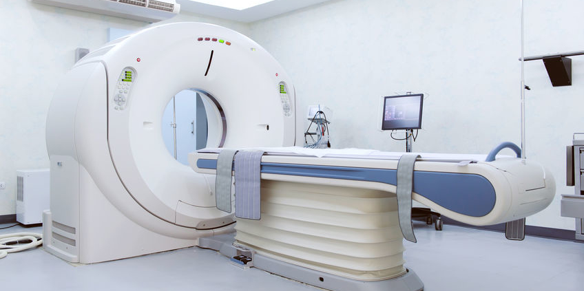
Breast MRIs are used in some newly diagnosed breast cancer patients, especially those with dense breast tissue. In addition, they are used as a supplement to mammography in women who are found to be at high risk. In order to distinguish between normal and cancerous tissue, an intravenous contrast dye is used. For MRI studies, the contrast material that is used is gadolinium-based.

Gadolinium is a heavy metal, and it has the potential to be retained in the brain and kidneys. Some recent studies in both the scientific and lay press have focused on the safety of gadolinium. While there have only been scattered reports of adverse events, an FDA advisory panel voted for warning labels and for more study.

The FDA advisory focuses primarily on a class of agents known as “linear”. The “macrocyclic” gadolinium contrast agents do not appear to have the same risk of depositing in the brain or kidneys. Both types of gadolinium contrast can be used for breast MRI studies. If you have concerns, ask the facility which form of gadolinium they use.

*Note – UCLA facilities (Westwood, Santa Monica and Santa Clarita), San Fernando Interventional Radiology (a RadNet facility) and Providence St. Joseph Hospital all use gadavist, a “macrocyclic” form of  gadolinium contrast, for their breast MRI studies. Because facility practice patterns may change over time, ask at the time of your exam if you have concerns.

8 May 2017

As a past-president of the American Society of Breast Surgeons I am probably more than a little biased. However, as always, the annual meeting held April 26-30th in Las Vegas was terrific. Topics including the full spectrum of breast disease, including benign and high risk lesions, genetic testing, breast cancer diagnosis and treatment including medical and radiation oncology updates, and metastatic disease.

The press briefing highlighted 3 abstracts which showed that:

  • Modern therapy for inflammatory breast cancer is associated with better outcomes than historically seen
  • Post-treatment lymphedema is related to a combination of treatments including surgery, radiation therapy, and chemotherapy – not just from surgery
  • Patients with DCIS have a 5 year risk of developing a cancer in the other breast of 2.8% and a 10 year risk of 5.6%, and patients should be discouraged from undergoing bilateral mastectomy for this condition. Developing a new cancer in the previously treated breast was twice as likely as developing a new cancer in the opposite breast, and the use of tamoxifen reduced the likelihood of any recurrence.

Dr. Nathalie Johnson moderated a pre-meeting course on Building a Breast Cancer Survivorship Program. I was invited to speak on Traditional Versus Virtual – Options for Patient Support and Education. Just as it can be challenging to choose between cake and ice cream (2 really good things), patients note advantages to both in person and online support and education. It doesn’t have to be one or the other – do what works for YOU! My slides are posted on SlideShare.

During the general sessions, a few topics stood out to me:

Dr. Shelley Hwang from Duke University spoke on DCIS subtyping and overtreatment. She noted that DCIS now comprises over 20% of all mammographically detected breast cancer. It is considered a “non-obligate precursor” of invasive cancer – the rate and likelihood of progression to invasive cancer are not clearly known. However, it is clear that some patients will never exhibit progression to invasive disease, and she discussed this in the context of thyroid and prostate cancer – two situations where we know that treatment in some patients will not provide the patient any benefit. The challenge is to sort out which patients will benefit from treatment and which ones will not. The COMET study is currently enrolling patients with low grade DCIS to in an attempt to help answer these questions.

Dr. Virginia Herrmann from Washington University in St. Louis spoke on non-genetic breast cancer risk factors. This is an important topic and I believe one that doesn’t get covered enough. She noted that hormone replacement therapy does increase risk – although the incremental risk is small and is seen only after about 5 years of use. However, longer term use does result in higher risk. Increased body mass index (BMI) is associated with risk – the risk of breast cancer is 30% higher in patients with a BMI greater than 31 kg/m2 compared to a BMI of 20 kg/m2. She noted that there is a linear relationship between alcohol intake and cancer risk, noting a 10% increase in risk for each 10 gm/day (for wine this is a little over 3 oz) increment in alcohol consumption. The risk is most associated with post-menopausal breast cancer, although in the study she quoted, only alcohol intake during age 50s was associated with an increased risk of postmenopausal breast cancer. She noted the association of ionizing radiation and breast cancer, and young women who received mantle (chest area) radiation for Hodgkin’s lymphoma have a markedly increased risk for developing breast cancer. She noted that breast cancer risk is increased in smokers, correlated with smoking intensity and duration. Finally, she noted the increased risk of breast cancer among soldiers stationed at Camp LeJune related to contaminated drinking water (tetrachloroethylene and trichloroethylene).

Dr. Tiffany Traina, a Memorial Sloan Kettering medical oncologist, gave a brief presentation about triple negative breast cancer: Searching For the Magic Bullet. There are several promising treatment strategies including targeting androgen receptors, the use of PARP-inhibitors in patients who have BRCA gene mutations, antibody-drug conjugates, immune modulating approaches, and targeted therapies based on tumor genomic profiles. Stay tuned – much more to come over the next few years related to this aggressive breast cancer subtype.

Dr. Lisa Newman, from the Henry Ford Health System in Detroit, spoke on Breast Cancer Outcomes: Disparities versus Biology. I have heard her speak on this topic multiple times over the years and always enjoy her excellent presentations. She noted that the incidence of breast cancer in black women is increasing, now close to that in white women. However, mortality rates for black women are higher than those for white women. There is an increased frequency of triple negative breast cancer in black women. She is involved in a research initiative evaluating the association between African ancestry and high risk breast cancer in white American women, African American women, and women in Ghana, including studying novel aspects of tumor biology and breast cancer stem cells – she is asking the question “are there differences in the oncogenic potential of mammary tissue that are associated with ancestry”? She concluded with what I felt was a powerful slide – 60% – 43% – 20%. Those were the survival rates for passengers on the Titanic who were in 1st – 2nd – 3rd class. She noted that healthcare outcomes are often dependent on access to care, and ended with a quote from Dr. Martin Luther King, Jr.: “Of all the forms of injustice, inequality in health care is the most shocking and inhumane”.

Dr. Stephen Edge, from the Roswell Park Cancer Institute, gave an update on the new American Joint Commission on Cancer staging system (AJCC 8th edition). Currently we stage breast cancer based on tumor size and lymph node status. However, it is recognized that that tumor biology plays an important role in prognosis and in some patients it may be more important that tumor size. The new staging system will incorporate tumor grade, Her2/neu status, ER/PR status, and Oncotype Dx status (if available) and should more accurately reflect prognosis. There are 422 lines in the new staging system – it will be impossible to memorize! Thankfully, he noted that the AJCC is working on a staging app.

The last day of the meeting held some great sessions, and the meeting room remained packed up until the very last minute. Dr. Ann Partridge from Dana Farber discussed special considerations in the young breast cancer patient. She noted that the disease is different, the patients are different, and the treatments should be different. Younger women have a higher likelihood to have more aggressive subtypes such as Her2/neu over-expressed and triple negative, and have lower survival rates than older women – even in those with the ER positive breast cancer. However, she cautioned not to over-treat patients based only on age. She noted that young age is not a contraindication for breast conservation, and that there is no clear improvement in mortality in patients who undergo more extensive surgery. She noted the need for improvements in treatment and support, including focused research and guidelines, which should lead to better outcomes.

Dr. Irene Wapnir from Stanford spoke on fertility preservation issues. She noted the various fertility options including medications and procedures. She also reviewed the POSITIVE trial, which will be assessing the risk of breast cancer relapse in patients who temporarily stop endocrine therapy to permit pregnancy, as well as to evaluate factors associated with successful pregnancy after interruption of endocrine therapy. She also stressed that fertility preservation should be discussed with any woman of childbearing age, whether or not she has had a prior pregnancy or a child – physicians won’t know what is important to their patients unless we ask!

Dr. Katherina Zabicki Calvillo from Dana Farber discussed breast cancer in pregnancy. She noted that 0.2-4.0% of breast cancers are diagnosed in pregnant patients – about 1 in 3000 pregnancies. She also noted that given the overall delay in childbearing (and the association of increasing age with breast cancer), the incidence of pregnancy-associated breast cancer will increase. Delays in diagnosis are related to hormonal changes which affect breast tissue making the exam more challenging, and that many patients and physicians assume that masses are related to pregnancy. She stressed that pregnancy termination is usually NOT required, but a multidisciplinary team approach is required. Many of these patients present in more advanced stages, but stage-for-stage, the prognosis is similar to non-pregnant patients with breast cancer. Chemotherapy can be given after the first trimester, but hormonal and Her2/neu targeted therapy should be avoided. She noted that mastectomy should be performed in the first and early 2nd trimester, and discussed the challenges of immediate reconstruction. Breast conservation could be considered in the late 2nd or 3rd trimester with post-lumpectomy radiation planned for after delivery.

Dr. Kevin Hughes from the Massachusetts General Hospital reviewed research studies that have found that in women over the age of 70 with early stage breast cancer, radiation therapy after lumpectomy may not be necessary.  The CALGB 9343 study showed that survival rates were the same whether women received radiation therapy or not. Radiation therapy did reduce the likelihood of cancer returning in the breast (local recurrence) from about 4% in the untreated patients to about 1% in the treated patients (after 5 years of follow up). However it is important to realize that the majority of women in that study were treated with endocrine therapy, which can help reduce the risk of local recurrence. As with many decisions regarding breast cancer treatment, a careful discussion of the risks and benefits of each option is necessary.

Dr. Tina Hieken from the Mayo Clinic gave a very interesting talk on the microbiome and the impact on breast cancer. We normally co-exist with many bacteria – we have ten times the more microbial cells compared to human cells. These microbes carry out metabolic reactions that can be essential to human health. The genetic material (genome) of our microorganisms is called the microbiome. She and her colleagues studied breast tissue from women with and without breast cancer and found that the background breast microbiome is different in women with breast cancer compared to those with benign conditions. She concluded by noting that the future may involve using a microbial pattern to predict breast cancer risk, exploiting the microbiome to enhance treatment response, and that there may also be implications for a cancer prevention vaccine. The Washington Post recently covered her research – definitely worth a read for more information.

Dr. Anthony Lucci from MD Anderson discussed the “Ongoing Saga of Circulating Tumor Cells”. We would all like to see the day when a blood test can tell us with certainty if cancer has developed or returned – but we’re not there yet. After reviewing several studies evaluating both circulating tumor cells (CTC) and circulating “cell free” DNA, he concluded that this information does provide prognostic information in both metastatic and non-metastatic patients, but is not in the current ASCO or NCCN guidelines for guiding treatment. Combining the CTC status with response to preoperative chemotherapy may identify a low risk subset of patients, but noted that additional studies are needed before we can reach the ultimate goal which is improving outcomes by monitoring and responding to CTC and cell free DNA levels.

Dr. Manjeet Chadha from Mount Sinai spoke on repeat lumpectomy after prior lumpectomy and breast radiation. Traditionally, mastectomy has been recommended if cancer returns after lumpectomy and radiation therapy. On average, there is about a 10% risk of “in breast” recurrence after lumpectomy and radiation, but this will vary based on tumor and treatment type. She reviewed several studies evaluating the different types of focused or partial breast radiation that may be used in selected patients who experience recurrence of their breast cancer. She also called for additional studies in this area.

One of the last talks was by Dr. Mehra Golshan from Dana Farber. He spoke about the decision whether or not to operate on patients with breast cancer who present with Stage IV (metastatic) disease. Traditionally, we have not recommended surgery for patients with metastatic breast cancer as these patients were not expected to have long survival, and it was not felt that removal of the main tumor would impact survival. Evaluating existing studies has also been challenging because while some have shown a benefit to removal of the main tumor, the patients who underwent surgery in those studies tended to be younger and healthier. He concluded by noting that surgery in patients with Stage IV breast cancer is not standard of care, but some studies do support this practice. It is recommended that these patients be evaluated in a multidisciplinary forum and that treatment choices be individualized.

 I returned from the meeting exhausted but energized. In addition to the scientific content, the meeting is an opportunity to connect with friends and colleagues across the country. I’m already looking forward to ASBrS 2018!

This post has not been endorsed by the American Society of Breast Surgeons.

23 August 2016

A study published in the Annals of Internal Medicine evaluated screening mammography taking into account breast density and breast cancer risk. For women age 50-74, the conclusion of the authors was that for women of average risk with low breast density (fatty or scattered fibroglandular), triennial (every 3 year) mammography screening averted the same number of breast cancer deaths as annual or biennial screening. Women screened every 3 years also had lower rates of biopsy procedures. For women at high risk with high breast density (heterogeneously or extremely dense), annual screening was better. High risk / high density patients accounted for approximately 1% of the study population.

The study was funded by the National Cancer Institute. The authors used simulation modeling which included national breast cancer incidence, breast density, and screening performance data. They did not include patients with genetic abnormalities such as BRCA 1/2 mutations. They also did not take into account the impact of MRI or tomosynthesis / 3D mammography.

Risk assessment involves a calculation (using various models) which takes into account a woman’s age, body mass index, menstrual and reproductive history, family history, prior biopsies, and other factors known to influence the risk of breast cancer development. In the current study, the authors used a risk calculator that takes into account breast density. Breast density is a factor associated with breast cancer, although studies vary regarding the impact of density on risk. Adding to the confusion, breast density rating is subjective – different radiologists may assign different density scores to the same patient. The model used in the current study also takes into account factors such as improved detection using digital mammography, improved treatment effectiveness, and the usual decrease in breast density that is seen with increasing age. It is unclear at this time which is the “best” risk assessment model to use – all have limitations, some significantly over-estimate risk, and none are a “crystal ball”.

So what should women do? The ideal screening test is one that is inexpensive, readily available and safe. It should also find cancers early enough to make a difference. Mammograms are an imperfect tool but they perform reasonably well in a wide variety of settings. The ideal screening program is to tailor the technology and screening frequency to the patient’s risk – one size never fits all. Women should be aware of their family history and risk factors, ask about their breast density, and then discuss these factors as well as their personal preferences regarding breast cancer screening with their physicians. True individualized and personalized risk-based screening is not yet a reality, but by making recommendations based on risk, we are taking steps in the right direction.

An Oklahoma mayor underwent a 3D mammogram (tomosynthesis) as part of a hospital promotion. She was diagnosed with ductal carcinoma in-situ (DCIS, also known as Stage 0 breast cancer) and she stated that the study “…saved my life.” She also recommended that women make sure to obtain a 3D mammogram. The story noted that tomosynthesis “virtually eliminates” the need for additional testing, and that early detection makes it less likely that the patient will need to undergo chemotherapy or radiation.

Here are a few errors in the story:

Statement: the mammogram saved my life
Fact: survival from breast cancer depends on many factors. The survival rate for patients with DCIS, regardless of treatment, is close to 97%. For invasive cancers, survival rates depend on stage as well as tumor biology. Small breast cancers “caught early” can still be lethal. It’s not the cancer in the breast that kills, it’s the cancer that gets to other areas of the body. Small tumors can and do spread.

Statement: early detection makes it less likely that a patient will need chemotherapy
Fact: the need for chemotherapy depends on tumor stage as well as tumor biology. As noted above, some very small, early stage breast cancers are very aggressive and have a high likelihood of spread, so chemotherapy is recommended. This is especially true for “triple negative” and “Her2/neu over-expressed” breast cancer subtypes.

Statement: early detection makes it less likely that a patient will need radiation therapy
Fact: radiation therapy is a standard recommendation for women with early stage breast cancer who undergo a lumpectomy. Since most women with early stage breast cancer are candidates for a lumpectomy, this statement simply doesn’t make any sense.

Statement: tomosynthesis “virtually eliminates” the need for additional imaging
Fact: while tomosynthesis can reduce the likelihood of needing additional views (“callback”) especially in women with dense breast tissue, diagnostic imaging with possible biopsy are still recommended when a concerning abnormality is seen.

While I certainly wish Ms. Noble well, stories like this always make me cringe, because they over-simplify a very complex situation. Here are some posts from Health News Review with some additional information:
Mayor: 3D Mammogram Saved My Life
3D Mammography and False Hope

27 June 2016

Choosing Wisely is an initiative of the American Board of Internal Medicine Foundation. Things Providers and Patients Should Question have been chosen by various medical societies based on review of the evidence-based literature. The American Society of Breast Surgeons (ASBrS) has published their contributions to the campaign (open access) in the Annals of Surgical Oncology.

The 5 ASBrS measures include:

1.) Don’t routinely order breast magnetic resonance imaging (MRI) in new breast cancer patients
2.) Don’t routinely excise all the lymph nodes underneath the arm in patients having partial breast removal (lumpectomy) for breast cancer when only one or two contain cancer
3.) Don’t routinely order specialized tumor gene testing in all new breast cancer patients
4.) Don’t routinely reoperate on patients if the cancer is close to the edge of the excised lumpectomy tissue
5.) Don’t routinely perform a double mastectomy in patients who have single breast with cancer.

Rational for the measures is included in the manuscript, and are posted on the Choosing Wisely website.

As with the other Choosing Wisely lists, these are meant to spark discussions between patients and their physicians regarding the appropriate use of medical tests and procedures. In many areas of medicine, tests and procedures may be performed without good supportive evidence. There may be many valid reasons for straying from an evidence based practice, which is part of the art of medicine. In fact, the Choosing Wisely website specifically states that “these recommendations should not be used to establish coverage decisions or exclusions.” In addition, the ASBrS statement notes that the selections made “are not meant to infer that a test or procedure endorsed in our list is a ‘never should occur’ event.”

Individualizing care as much as possible is extremely important. However, that does not mean performing every available test just because it’s available. Patients and physicians need to realize that there are downsides to unnecessary testing. Physicians also need to remain educated about current practice, and not continue to make outdated recommendations simply because they may have “always done it that way.” A goal of the Choosing Wisely campaign is to ensure that when decisions are made, available evidence is considered and that the process is a collaborative one between patients and their physicians.

26 April 2016

A growing number of states have enacted so-called “dense breast” legislation – requirements that radiology facilities inform patients if they have dense breast tissue, which may limit the sensitivity of mammography.

A study published in the Journal of the American Medical Association evaluated the dense breast legislation for 23 states who have these requirements. They evaluated the content, readability, and understandability of the dense breast notification. The authors found that the readability of these notices was at the high school level or above, and many notices had poor understandability. Also concerning was the fact that many notifications lacked concordance with the states’ average literacy.

Unless women (and their physicians) can make sense of the notifications, they are worthless. Research studies generally recommend that informed consents and study descriptions be presented to patients at a 6th-8th grade reading level, but these standards do not exist for other patient information.

It is important for patients to ask if there is anything about their medical test results or instructions that they do not understand.

17 January 2016

In October 2015, the American Cancer Society (ACS) updated their screening mammography guidelines, raising a lot of questions and concerns as annual mammograms were no longer recommended for all women starting at age 40. The United States Preventative Services Task Force (USPSTF) released new guidelines on screening mammography last week. The new USPSTF guidelines recommend an individualized risk-based approach for women age 40-49, and mammographic screening every other year for women age 50-74. The task force acknowledged that screening mammography reduces the likelihood of dying from breast cancer, but noted that the benefit was the greatest for women age 60-69, and the benefit was small for women age 40-49. For younger women, they noted that screening mammography is associated with higher false positives (false alarms).

The conflicting guidelines published by the USPSTF, ACS, and other organizations have caused much confusion. Many women simply want to know what they should do. The reality is, screening mammography is a one size fits all tool, and as any woman knows, that doesn’t really work. The sensitivity (“effectiveness”) of mammograms will vary depending on a patient’s age, menstrual status, use of hormone therapy, body mass index, breast density, and other factors. It is unrealistic to expect that one test can account for all of those variables, as well as personal risk factors for breast cancer, and personal preferences regarding screening risks and benefits.

To state it very simply:

  • Mammograms may lead to improved survival from breast cancer by detecting some cancers early
  • Some breast cancers are so aggressive that early detection does not lead to improved survival
  • Mammograms are imperfect

It is important to note that the USPSTF and ACS recommendations apply to average risk women. Unfortunately, risk assessment is not routinely performed, and many factors associated with an increased risk, such as heavy alcohol intake and breast density, are not taken into account in the existing models. In an ideal world, we would swab a cheek, obtain some DNA, and then determine whether or not a woman is predisposed to develop breast cancer [I’m thinking beyond BRCA and other genetic testing here]. If we can determine that a 30 year old woman, even without a family of breast cancer, is predisposed to develop the disease, we can make educated screening recommendations (perhaps ultrasound and/or MRI, as mammograms are not very helpful in very young women).  If we can determine that a woman is highly likely to die of a heart attack by age 60, and has no cancer predisposition, she might make the decision to forego screening mammography. Unfortunately we’re not quite there yet, but there are genetic risk tests under development.

I recently spoke with a researcher specializing in cancer risk. We discussed that at the end of the day, multiple organizations will continue to look at the evidence and draw different conclusions, so in his opinion, it wasn’t realistic that we should expect consensus. Physicians, researchers, and organizations are currently spending a lot of time and effort arguing with each other about whether mammography should start at age 40, if mammograms should be performed every versus every other year, and when screening should stop. However, our time and efforts might be better spent by working together to address issues such as disparities in access to screening mammography and lack of access to quality mammography and breast care for many women.

Recognizing the variability of each woman and her breasts, acknowledging the uncertainties regarding the risks and benefits of screening, and focusing on a shared decision making approach might help reduce some of the confusion. In medicine, as in many areas in life, there is often more than one right answer. The same applies to screening mammography. Time to move beyond the one size fits all approach.

Additional Reading:
New York Times: Panel Reasserts Mammogram Advice
JAMA: A Public Health Framework for Screening Mammography: Evidence-Based vs Politically Mandated Care 
New York Times: Insurer Rewards Push Women Towards Mammograms
Forbes: A Turning Point in the Breast Cancer Screening Debate?
NPR Shots: Federal Panel Finalizes Mammogram Advice That Stirred Controversy
OncLive Final USPSTF Guidelines
Cornell Mammogram Decision Aid for Women age 40-49