6 March 2019

The American Society of Breast Surgeons (ASBrS) held their annual meeting in Dallas last week. This meeting usually draws about 1500 breast surgeons (just under half the ASBrS membership) from around the world, for several days of pre-meeting courses, didactic sessions, and research presentations. In addition to the science, the meeting provides opportunities for breast surgeons in all types of practice settings and at all levels of training and practice to network and learn from each other. 

The following covers some highlights from the general session. 

The meeting started off with the Critical Issues in Breast Cancer Forum: Changing Paradigms for Breast Cancer Surgery. Dr. Cary Kaufman presented an update on current clinical trials for cryoablation for breast cancer. Cryoablation is a technique that freezes the tumor, using a small probe placed into the tumor (similar to a needle biopsy) under local anesthesia. There are several types of ablative therapy including laser, radiofrequency, high-frequency ultrasound, and cryoablation. Because cold is a natural anesthetic agent, patients undergoing cryoablation do not need any sedation, and the procedure is performed while they are awake. 

Cryoablation was initially tried with benign tumors (fibroadenomas). In many cases, the fibroadenoma reabsorbed, leaving no mass and only a tiny (3 millimeter) scar. Multiple studies have looked at the use of cryoablation for breast cancer, and most have restricted therapy to patients with small (1.5 cm or smaller) estrogen receptor positive, Her2/neu negative tumors. I participated in a national multi-center trial, the ACOSOG / ALLIANCE Z1072 trial, which was published in 2016 and demonstrated that cryoablation was successful in the majority of these patients. All patients in the ACOSOG / ALLIANCE Z1072 study underwent surgery within one month of the ablation, so that the tumor site could be removed and evaluated. Several subsequent studies have looked at cryoablation for breast cancer without surgery. The longest follow up was from Dr. Fukuma in Japan. After 12 years of follow up, he reported 3 local (in-breast) recurrences in 304 patients. Combining 3 published trials, Dr. Kaufman noted that local recurrence rates range from 0.98 – 1.4%, and he concluded that this is extremely promising technology. He also noted that cryoablation of breast cancer appears to have an immunologic benefit – when the tumor cell membranes are disrupted by the extreme cold, the patient is exposed to tumor antigens, which may prompt antibody formation. It is very premature to determine if this immunologic effect will help reduce recurrence rates.

Dr. William Small presented updates on 3 clinical trials of intraoperative radiation therapy (IORT). An advantage of IORT is that it is delivered at the time of lumpectomy, in the operating room, as a one-time treatment. A disadvantage is that status of the lumpectomy specimen margin and lymph nodes are not known at that time. If it is found on final pathology that there are positive margins, external beam radiation is recommended, and at least one trial noted that approximately 30% of patients who received IORT required additional whole breast radiation. Most studies of IORT have been limited to “low risk” lesions – small, low grade invasive cancers in older women. He discussed that a criticism of these studies is that some of these women may not have needed radiation therapy at all. Dr. Small noted that local recurrence rates are slightly higher (3.3 versus 1.3%) but that statistically, IORT is considered “non-inferior” to whole breast irradiation. He noted that seroma (fluid accumulation) is more common in patients who undergo IORT.  He concluded by stating that there is an “acceptable” toxicity, with non-inferior local recurrence. However, as there is relatively short follow up available in low risk patients, he questioned the applicability of this procedure to a broader patient population. A US registry is planned.

Dr. Antonio Toesca presented the results of his study of 100 patients who underwent robotic nipple sparing mastectomy (NSM) and implant reconstruction, and showed a fascinating video which highlighted the precise and meticulous dissection, along with improved visualization, compared to a standard surgical procedure. The average incision size was a little over 1 inch, and the specimen was removed intact (in one piece). The procedure averaged 1 hour and 18 minutes longer than their standard for a nipple sparing mastectomy and implant reconstruction (3 hours, 36 minutes for the robotic procedure. Patients who underwent the robotic procedure were less likely to have axillary web syndrome and reported Improved physical, psychological and sexual well-being. 

Why could performance of NSM using robotic technology become important? Dr. Tina Hieken presented the results of her study (abstract 580759, page 31) showing that as experience with the procedure has grown, indications are expanding and patients who previously were not candidates for the procedure are now being considered. A NSM is a technically challenging procedure, and it takes a toll on the neck and back of a surgeon. A 2017 study published in JAMA Surgery noted a high incidence of work-related musculoskeletal disorders among surgeons and interventionists. Dr. Katherine Kopkash presented her research (abstract 51837, page 52) using intraoperative electromyography (EMG) on the surgeon to assess muscle strain during NSM. Of course, oncologic safety is the primary concern, and more study on the long-term outcomes (as well as costs) of robotic procedures is required. 

The next session was Emerging Strategies in Breast Cancer Care, which focused on “de-escalation” of surgical therapy. Dr. Anna Weiss provided an update of clinical trials evaluating active surveillance for low-risk ductal carcinoma in-situ (DCIS): COMET, LORD and LORIS. Approximately 60,000 cases of DCIS are diagnosed annually. Patients undergoing active surveillance do not have surgery, some are treated with endocrine therapy, and all undergo regular monitoring. This is a accepted option in select cases of prostate cancer, and Dr. Weiss noted that there is no difference in overall survival in patients with low-grade DCIS who do not undergo treatment. The LORD and LORIS trials are open in the UK and the COMET study is open in the US. (Additional perspective)

Dr. Henry Kuerer presented his research on the percutaneous management of breast cancer in the setting of a pathology complete response (pCR) following neoaduvant (before surgery) chemotherapy. He noted that for survival and recurrence matter most, but side effects and complications are significant concerns for both patients and physicians. I’ve recently covered details of his research on this blog

Some of the twitter conversation related to this talk included patients who noted that they would rather undergo surgery than chemotherapy. It is important to note that the patients involved in this study are those who were going to be treated with chemotherapy regardless of surgical therapy because they have triple negative or Her2/neu positive breast cancer. In these patients, systemic (whole-body) therapy is necessary due to the higher likelihood of metastatic disease. Surgical therapy in these patients, especially the “exceptional responders”, may not improve outcomes, but of course more study is needed. Surgery remains the standard of care for breast cancer therapy.

Dr. Judy Boughey discussed several cooperative group trials evaluating management of the axillary (underarm) lymph nodes, and these studies are also focusing on how we can safely de-escalate axillary surgical therapy after neoadjuvant chemotherapy. This is an area that is rapidly evolving with expansion of the criteria for a less aggressive approach to the axilla.

In a session on Evidence-Based Prevention and Management of Surgical Complications, Dr. Suzanne Klimberg presented on chronic post-mastectomy seroma. A seroma is a fluid collection – fluid normally accumulates after mastectomy which is why drainage tubes are left in place. Normally, drains can be removed after 7-14 days, but about 30% of patients will develop prolonged drainage. This is a frustrating problem for patients and physicians as the persistent fluid can be uncomfortable, may increase the risk of infection, and may delay the start of planned chemotherapy or radiation. She noted that a surgical technique to close the tissue known as “quilting” can reduce the rate of chronic seroma, but that it results in excessive skin dimpling and has a significant impact on the cosmetic results. She stated that additional drainage tubes, various “sealant” agents and compression (such as wearing an ace wrap) are not effective. The area may be sclerosed (scarred) by instilling talc or antibiotics, and in some cases, re-operation to remove the inflamed tissue is indicated. Otherwise she recommended patience and repeat aspirations. She noted that there are no ways to successfully prevent seromas from forming.

Dr. Amal Khoury presented on chronic post-mastectomy pain, and noted that persistent pain occurs in 25-60% of patients undergoing any type of breast surgery. It is thought that this chronic and at times severe pain is due to damage to and neuroma formation of the cutaneous (skin) branches of nerves that run along the 4thand 5thribs, which are roughly at the inframammary fold (bra line below the breast). These cutaneous nerve branches are often not visible at the time of surgery. She noted that the pain syndrome it is often not recognized, and when recognized it is often not treated effectively. She stated that injections with a combination of long-acting local anesthetic and steroid (in a very small dose) at the trigger points is more effective than taking pain or other medications, and in their study at UCSF, 91% of patients required only one injection for lasting relief.

The next session was Practical Considerations for Systemic Treatment. Dr. Judy Boughey reviewed the I-SPY2 clinical trials, which utilize an innovative “adaptive randomization” approach in patients who are undergoing neoadjuvant chemotherapy for triple negative, Her2/neu positive, or other high risk breast cancers. pCR rates are assessed, and drugs that are successful move up higher in the randomization algorithm. This study and its flexible randomization protocol have accelerated the use of some novel agents. Patient reported outcomes assessing quality of life, fear of recurrence, symptoms and side effects are being assessed. If drug response rates are similar, the “winner” may be the one associated with fewer side effects. Dr. Barry Rosen discussed specific strategies to identify the previously involved axillary lymph nodes when chemotherapy is performed prior to surgery. Dr. Elizabeth Mittendorf presented on breast cancer immunotherapy and surgical implications of these treatments. She noted that one agent, atezolizumab, is currently approved for use in patients with metastatic triple negative breast cancer. She noted that there are concerns about wound healing complications with these agents but unfortunately the clinical trials did not specifically assess for this. In addition, she noted that some immunotherapy agents are associated with development of adrenal insufficiency – this complication has only been reported in a small percentage of patients, but it is an important consideration in any patient who is going to have surgery.

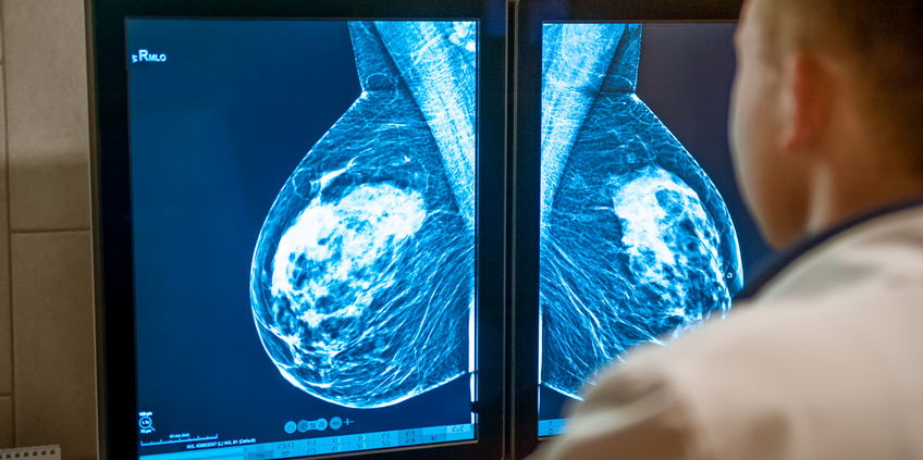
A session was held on breast imaging. Dr. Molly Sebastian presented on the impact of breast density on breast cancer risk, noting that it is more difficult to screen patients with dense breasts, and that these patients are also at increased risk for developing breast cancer. The associated breast cancer risk increases with the level of density. Approximately 50% of women in US are considered to have dense breast by mammogram, and she cited a 2010 study that found that 30% of breast cancers could be linked to highly dense breast tissue. Contributors to increased density include younger age, use of hormone replacement therapy, race (Asian), diet (Western), alcohol use, and hereditary factors. She did stress that the presence of a germline genetic mutation (such as BRCA 1/2) conveys a much higher level of risk (regardless of density) than breast density itself. 

Dr. Brigid Killelea discussed balancing high-risk screening (which usually includes MRI) with the concerns about gadolinium toxicity. Gadolinium is a “rare earth heavy metal”, and is used in the contrast material that is administered (using an intravenous line) when breast MRI is performed. Acute allergic reactions are uncommon but as gadolinium is excreted through the kidneys, there are concerns about the potential for kidney damage especially in patients with pre-existing renal insufficiency. Nephrogenic systemic fibrosis (NSF) is an unusual condition that results in progressive deposition of gadolinium in the skin. It has also been found that the number of exposures to the linear form of gadolinium (as opposed to macrocyclic, which is what is most commonly used with breast MRI) correlates with increasing deposits in the brain. More research is needed to determine if this leads to an increased risk of Parkinson’s or other diseases. Studies evaluating “fast” MRI protocols are ongoing but they still use gadolinium contrast. Some work is being done with non-contrast MRI and Dr. Killelea noted that it shows some promise in detecting certain lesions. 

In the session on Ethical Issues in Breast Cancer Surgery, Dr. Rachel Greenup discussed how to manage the situation when the principles of respect for patient autonomy conflict with the standard of care. She noted that patient autonomy allows for us (as physicians) to educate but not to decide care for patients, and that poor physician-patient communication is a key factor in patients opting for non-standard care. Factors associated with patients declining standard therapy include a negative first experience, an uncaring / insensitive / unnecessarily harsh oncologist, fear of side effects, and belief in the efficacy of alternative therapy.  In regards to endocrine therapy for breast cancer, she noted that unmanaged side effects are a significant contributor to stopping therapy. She also presented data showing poorer outcomes in patients who declined standard therapy, and that many, when faced with disease progression, did then opt for conventional treatment. She recommended that physicians review and present evidence to their patients in an understandable way, taking time to acknowledge fears and address patient barriers to treatment, provide time to adjust to diagnosis, suggest a 2ndopinion, and avoid abandonment or fear tactics. She also suggested that physicians be more open (when medically safe) to the combination of alternative and standard therapy. She stressed that patient autonomy is the priority, and that open communication can help align patient-centered care with evidence-based care. 

Dr. Terry Sarantou discussed the ethical issues of obtaining informed consent when performing a new surgical procedure, noting that there is FDA oversight for new drugs and surgical devices, but not for surgical procedures. He stressed that informed consent is a communication process, not a form to be signed. 

Recognizing the role that surgeons play in the current opioid crisis, Dr. Sarah DeSnyder discussed proper prescribing of narcotics in breast surgery. There was also an abstract presentation by Dr. Betty Fan (abstract 5808940, page 27) on this subject. She noted that women who expected postoperative pain or those who reported higher preoperative distress used more postoperative opioids for pain management. She stressed that physician and trainee education about proper prescribing is critical as is setting patient expectations for postoperative pain and providing non-narcotic options. The use of nerve blocks, long-acting local anesthetic agents, acetaminophen (Tylenol) and ibuprofen were also discussed. 

Photographs are an important part of breast and reconstructive surgery to document results both for patient and physician education as well as for quality assurance, and Dr. Toan Nguyen reviewed some of the ethical, legal and technical considerations to protect patient confidentiality and privacy. The ASBrS statement on this issue has been published in the Annals of Surgical Oncology.

In the session covering New Perspectives on Old Problems, Dr. Lee Wilke noted that with improved surgical techniques, breast conservation is now appropriate for select patients with more than one tumor in the breast. She did note that in up to 20-30% of patients with more than one tumor in the breast, the tumors are different subtypes, which may have implications for therapy – so pathologic analysis needs to be performed on all lesions. Dr. Stephen Grobmyer reviewed the current literature on local (in-breast) recurrence, noting that repeat breast conservation may be appropriate in some patients. However, if repeat radiation is performed, there is a higher risk of skin toxicity and potentially unacceptable cosmetic results. In addition, for left-sided breast cancers, repeat radiation raises concerns about cumulative radiation damage to the heart. Repeat lumpectomy without radiation is associated with a 20-40% risk of local recurrence. IORT may be utilized in some patients, but studies are ongoing and data is limited.

Dr. David Euhus discussed that genetic testing does not only potentially impact the surgical procedure that is recommended, but may influence the decision for radiation therapy as well as systemic therapy. In addition, results of genetic testing may impact surveillance for additional breast or other cancers in the patient as well as recommendations for family members. The ASBrS recently updated their genetic testing guideline, recommending that genetic testing be considered for newly diagnosed breast cancer patients. (Additional perspective)

In the session on Benign Breast Disease, Dr. Jane Mendez reviewed breast fistulas (persistent drainage through the skin) and infections, and Dr. Vincent Reid reviewed some of the non-malignant masses that can develop in the male breast. Dr. Katrina Mitchell, who is a breast surgeon as well as a certified lactation consultant, provided recommendations for management of post-partum patients who develop mastitis or breast abscess. One of the key recommendations was that patients should continue breast feeding (better than pumping for keeping the breast empty) and that patients do not need to “pump and dump” the milk while on antibiotics. 

Dr. Stephanie Valente discussed breast pain, which is a common problem that frustrates both patients and physicians. Pain is a symptom of breast cancer in less than 2% of cases.  Suggestions for treatment include decrease caffeine, nicotine, and dietary fat intake, and consider supplementation with essential fatty acids such as evening primrose oil (EPO) or vitamin E. However, she noted that that some studies show that EPO and vitamin E are no better than placebo. Both flaxseed and chasteberry have shown to be effective. Diclofenac (a non-narcotic pain medication) gel can be effective but it needs to be used for several weeks before improvement is seen and it is expensive. In severe cases, danazol (an androgen hormone) or tamoxifen can be used but are associated with significant side effects.

There were several sessions on oncoplastic surgery. Oncoplastics refers to combining oncologic (cancer) surgery with attention to cosmetic outcomes. Basic principles include placing the incision in the least conspicuous place and closure of as much of the breast tissue once the tumor has been removed as possible to minimize, or preferably avoid, a depression in the area. More advanced techniques include rotation flaps and mastopexy (lift) that may be performed by breast surgeons or breast surgeons collaborating with their plastic surgical colleagues. There was also a session discussing some of the advanced microvascular procedures that are being studied to treat lymphedema as well as a video session showing some basic techniques to perform a better (flat) closure for patients undergoing mastectomy without reconstruction. 

The keynote address was delivered by the actress Kathy Bates. Ms. Bates underwent a bilateral mastectomy for breast cancer and has bilateral arm lymphedema. She is a spokeswoman for the Lymphatic Education and Research Network, working to educate, support, and advocate for patients who have lymphedema. She delivered a moving and unique address to the group, combining science and her personal patient perspective. An abstract (abstract 581304, page 22) presented during the meeting demonstrated that postoperative surveillance with bioimpedence spectroscopy compared to tape measure resulted in a 10% decrease in the number of patients requiring complex decongestive physiotherapy. However, these results, which were a planned interim data analysis, did not reach statistical significance.

The new ASBrS screening mammography guidelines were released at the meeting. They recommend formal risk assessment starting at age 25 and a risk-based approach to screening, as well as annual mammography starting at age 40 for average-risk women. (Additional commentary)

All of the research abstracts and posters can be found here. There were many interesting and thought-providing presentations, but it is important to remember that abstracts represent incomplete data and have not been subject to the peer-review process. The oral abstracts that were presented will be published in manuscript form later this year. The poster gallery can be found here (not all posters have been uploaded by the presenters).

As usual if anyone is interested in one of the articles referenced but does not have access, or wants additional information, please send your email address to me: contact at drattai dot com and I will be happy to respond.

This post has not been endorsed by the American Society of Breast Surgeons.

3 May 2019

The American Society of Breast Surgeons (ASBrS) has updated their screening mammography guideline, recommending that women of average risk begin annual screening mammograms at age 40. This update brings the ASBrS guideline more in line with those of the American College of Radiology and the Society of Breast Imaging, and are a departure from their previous guideline, which called for screening to start at age 45. The American Cancer Society recommends annual screening from age 45-54, followed by biennial screening with the opportunity for annual screening. The US Preventative Services Task Force recommends biennial screening starting at age 50.

The updated ASBrS guidelines recommend as a first step that all women undergo a formal risk assessment starting at age 25. Risk assessment involves using any one of a number of models to estimate 5-year and lifetime risk of breast cancer development. These models take into account age, family history, menstrual and pregnancy history as well as history of prior biopsy and racial / ethnic background. Some models take into account body mass index and breast density. The ASBrS guideline has additional recommendations, outlined below, depending on risk level. The recommendations listed for women at high risk of breast cancer are consistent with those of the American Cancer Society and National Comprehensive Cancer Network.

When to start and how often to perform screening mammography has been the subject of much debate over the years. The primary issue is the number of lives saved balanced against the harms of screening mammography. Harms include false negatives (cancer is not detected by the mammogram), false positives (suspicious areas that turn out to be benign) and recommendations for additional imaging (added cost and concerns about the potential effects of cumulative radiation). There have also been concerns raised about over-diagnosis and over-treatment: finding cancers that would never pose a threat to a woman’s health or life – but the patient is exposed to the potential harms of cancer therapy.

The goal of screening is not to detect all cancers – otherwise we would recommend complete body imaging for everyone. The goal of screening is to improve the survival from the cancers that are detected. An ideal screening test is relatively inexpensive and performs equally well in the patient population undergoing the screening. Mammography is acknowledged to be an imperfect screening tool – while relatively inexpensive and safe, it simply does not perform the same in women with breasts of different ages and densities. Approximately 1000 women need to be screened to detect 2 – 7 breast cancers, and mammography performs best in women age 50-74. In younger women, a few cancers will be detected, and there may be a survival benefit. However, because breast cancer is less common in younger women, a larger number of women need to be screened to find a single cancer, increasing the likelihood of a harm. The benefits of screening do not always outweigh the risks – at least for an individual patient.

It is also important to note that some of the new ASBrS recommendations, such as 3D mammography as the preferred type of mammogram, supplemental ultrasound in women with dense breasts, and MRI for all women with a history of breast cancer, are still undergoing investigation and are not always covered by insurance. We know that these studies will find additional cancers, but we do not yet have data on improved outcomes. My patients know that I am fond of saying “the more we look, the more we find, but not everything we find needs to be found.”

Two ongoing studies will hopefully provide additional information regarding 3D mammography and screening interval:

  • The TMIST trial is a national multi-center study assessing the performance of 3D compared to 2D digital mammography. This study will assess rates of cancer detection, callbacks and benign biopsies as well as biology of the tumors detected and outcomes.
  • The WISDOM study is evaluating a risk-based screening approach. After a comprehensive risk assessment, genetic testing, and assessment of risk tolerance, patients are assigned annual versus biennial screening. Women can self-enroll in this study (disclosure – I am not involved with the study team but I am enrolled in the study as a participant) and do not have to change mammography facility.

Until we are at a point when an individual woman’s level of risk can be accurately predicted, there is no definitive “best” screening guideline to follow. Recognizing the potential harms of screening mammography, it is no longer appropriate for physicians to simply hand out an order for an annual screening mammogram for all women starting at age 40. A balanced discussion, taking into account an individual woman’s risk and level of risk tolerance as well as the absolute potential benefits and harms of mammography is indicated. These discussions are nuanced and take time. Various genomic assays are being evaluated and it is hopeful that we will at some point have a test that can accurately predict a woman’s risk of breast cancer – which can then be used to provide more tailored guidance regarding imaging. Until that time (and I hate to end this way…), talk to your physician and medical team about what screening option is right for you.

7 April 2019

The Society of Surgical Oncology held their annual meeting in San Diego, CA from March 27-30, 2019. Approximately 1700 surgical oncologists were in attendance. As the organization is geared towards the entire field of surgical oncology, only a portion of the meeting covered breast cancer. Here are some of the highlights:

Genetic Testing and Management
Dr. Judy Garber – Dana Farber
Updates in Testing and Management of BRCA Mutations
BRCA Mutation information from the National Cancer Institute
– Consider repeat testing if original genetic testing was performed prior to 2012 as more genes as well as pathogenic mutations have been discovered
– NCCN guidelines for breast cancer surveillance in BRCA 1/2 mutation carriers:
o Clinical breast exam every 6-12 months starting at age 25
o Annual MRI age 25-75 (individualize after age 75)
o Annual mammogram age 30-75 (individualize after age 75)
– NCCN guidelines for breast cancer prevention in BRCA 1/2 mutation carriers: discuss mastectomy, discuss tamoxifen
– Premenopausal BRCA mutation carriers who undergo oophorectomy experience breast cancer risk reduction. The level of breast cancer risk reduction in BRCA1 carriers is lower than in BRCA2 carriers as BRCA1-associated tumors are more likely to be triple negative
– Prenatal genetic testing is available in mutation carriers, and may be used for selective reproduction
– BRCA 1/2 mutation status does not impact breast cancer outcomes; tumor biology impact on outcomes is independent of mutation status
– BRCA 1/2 are DNA repair genes. Tumors associated with BRCA 1 tend to be triple negative and tumors associated with BRCA 2 tend to be ER/PR+, Her2- (but all combinations have been seen)
– Clinical trials are evaluating the use of cisplatin chemotherapy in patients with BRCA mutations – cancer cells are not able to repair DNA-induced chemotherapy damage due to the defective BRCA gene
– PARP inhibitors interfere with DNA repair and have traditionally been used to treat ovarian cancer. Small studies show some effect in breast cancer in the setting of BRCA mutations. Larger studies are ongoing. So far they only seem to work in breast cancer when there are BRCA mutations
– A challenge to treatment with PARP inhibitors is that there are many mechanisms of resistance, and tumors demonstrate a variable response to therapy – tests are being developed to predict response
– Lurbinectedin – a drug from sea slugs (!) may have some effect
– A very interesting comment – Dr. Garber noted that DNA breaks may be immunogenic, so there may be a role to combine PARP inhibitors and immunotherapy treatments
– Denosumab, a RANK-ligand used for bone protection in breast cancer patients, may have breast cancer risk-reducing activity – a randomized trial is pending to assess its activity as a preventative agent

Thuy Vu, Genetic Counselor – Wake Forest
What Genetic Test Should I Order?
– Once the appropriate patient for genetic testing has been identified, how to decide what lab to use? Consider lab experience, as well as cost and insurance support
– Patients with a complicated family history (multiple different cancers in scattered relatives), absent family history (adopted), and evidence of multiple cancer syndromes will benefit from NGS (next-generation sequencing) genetic panel testing
– A disadvantage of broad genetic panel testing is that there is currently incomplete information on all of the mutations that may be identified. Risk for cancers unrelated to the current diagnosis may be identified. In addition, there will be an increased prevalence of variants of uncertain significance (VUS)
– She noted to use caution when patients bring in test results from ancestry.com and similar companies – these sites often assess for SNPs (single nucleotide polymorphisms), which is not the same as testing for a genetic mutation, and full genetic testing may need to be repeated
– She acknowledged that there is a shortage of genetic counselors, even in large university centers. Many testing companies and labs now have associated genetic counselors, and there are some independent companies offering telephone counseling services

Dr. David Euhus – Johns Hopkins
ATM, CHEK2 and Other Genes
– While multiple gene mutations influencing breast cancer risk have been identified, they do not all convey the same level of risk
– As testing for multiple genes has increased, BRCA mutations are no longer the most common mutations found
– High risk genes include BRCA 1/2, TP53, PTEN, PALB2, STK11, CDH1
– Moderate risk genes include ATM, CHEK2, NBN, NF1
– These and other genes explain approximately 14-28% of genetic risk for breast cancer – most patients with a strong family history of breast cancer do not have an identifiable mutation
– There is a range of risk associated with all of the genes that in part depends on the mutation type – what type of damage does the mutation cause to the DNA. Family history of breast cancer can modify risk.
– For most of these patients, NCCN guidelines recommend annual MRI in addition to mammograms. Age to start supplemental screening depends on the mutation.
– He noted that increased screening for other associated cancers when there is no clinical benefit leads to patient harms – financial, emotional, and physical
– A good question from the floor about the role of ultrasound as supplemental screening (in addition to MRI) – Dr. Euhus states he uses 3D mammogram / tomosynthesis and does not use ultrasound unless the patient is pregnant / lactating

Dr. Kevin Hughes – Massachusetts General Hospital
What the Surgeon Needs to Know about Genetic Testing
– High cost of testing is not the problem – interpretation of the results is the challenge
– Assuming that approximately 10% of breast cancers are hereditary, over 51,000 breast cancers could have been prevented with testing
– For the breast surgeon, understanding BRCA 1/2 is not enough. There are many genes, each have different spectrum of associated cancers and associated risk; treatment needs to be individualized for the patient taking into account their specific mutation and family history
– He emphasized the point Dr. Garber made that if testing on a breast cancer survivor was performed prior to 2012, those patients should be re-tested
– Recent American Society of Breast Surgeons guidelines call for consideration of genetic testing in all breast cancer patients
– Dr. Hughes notes that this is already a standard recommendation for other cancers such as ovarian, pancreas and others
– The field is becoming more complicated – it is not expected that anyone can memorize this – go to the internet and look it up!

Resources:
ASK2ME – All Syndromes Known to Man Evaluator
ClinVar – look up specific mutations to see how they have been classified
PROMPT registry for patients with rare mutations

Breast Cancer Treatments in the Young and Elderly
Dr. Mina Sedrak – City of Hope
Treatment Strategies in Octogenarians with Early Stage, High-Risk Breast Cancer
– Incidence and mortality from breast cancer increase with age; the number of older adults in the US is increasing
– Breast cancer outcomes are often worse for older (as well as younger) women
– Older adults are underrepresented in cancer clinical trials – 1/3 of patients with breast cancer are over the age of 70, but only a small percentage of them are included in clinical trials
– Because of lack of clinical trial data in older women, patients may be under- or over-treated [DJA note – we have a similar situation in men with breast cancer].
– There is no universal definition of “old”. Aging is a continuous spectrum, and chronological age does not accurately predict functional age. The ASCO Guidelines Geriatric Assessment can help understand factors other than chronological age to predict morbidity and mortality. US Life Tables can also be used to estimate life expectancy, as well as ePrognosis. Estimation of life expectancy should be performed for all older patients before making a treatment plan
– How to best treat cancer in the elderly patient: it depends on life expectancy, aging concerns, risks / benefits of treatment and the potential impact of co-existing medical problems
– What risks can we modify and what are the patient preferences? There is no “one size fits all”

Dr. Tyler Chesney – University of Toronto
Adjuvant Radiotherapy for Older Women after Breast Conserving Surgery
– 4 randomized clinical trials addressed if elderly patients with low-risk breast cancer need radiation therapy after breast conserving therapy: NSABP B-21, A. Fyles, CALGB 9343, and PRIME II studies
o Meta-analysis of these 4 studies: 2387 patients across all trials, early stage breast cancer, hormone receptor positive. Addition of radiation therapy reduces local recurrence from 60 versus 10 / 1000 at 5 years. 2 trials had 10 year follow up, noting recurrence was 80 versus 20 / 1000 women.
o 3 of the trials provided data on axillary recurrence: absolute benefit was small, 12 versus 3 / 1000 women. No difference in distant recurrence or overall survival
– Prime I study showed that older women who underwent radiation therapy had increased fatigue over 5-10 years but similar overall health-related quality of life
– Accelerated partial breast irradiation may be an option, but some studies have shown higher local recurrence and poorer cosmetic result (depending on treatment method)
– While toxicities of radiation therapy have improved with more modern techniques, logistical concerns such as time, need to travel, and cost may be of higher concern for older women

Dr. Laura Dominici – Dana Farber Cancer Institute
Reconstruction and Body Image in Young Patients
– More than 13,000 women under the age of 40 are diagnosed with breast cancer annually in the US, approximately 7% of all new diagnoses
– Younger women newly diagnosed with breast cancer have been shown to have higher rates of anxiety and distress after diagnosis, they have historically received more aggressive treatment, and have a long survivorship period
– More aggressive surgery such as mastectomy does not lead to improved overall or breast cancer specific survival. Local recurrence is related to tumor biology, not age of the patient
– Mastectomy (single and bilateral) rates are rising, especially among younger women. Rates of reconstruction are increasing, as are rates of post mastectomy radiation
– A growing number of patients are “going flat” after mastectomy, opting for no reconstruction
– Dana Farber young women’s multicenter prospective cohort study: poorer satisfaction with breast-related, psychosocial and sexual well-being after unilateral and bilateral mastectomy. Other factors impacting poorer satisfaction include financial status, lymphedema, and the need for radiation
– 42% of women age 50 and younger (in the Dana Farber study) regret their surgical decision including primary surgery and reconstruction decision. Patients in this study were not asked what the actual regret was – doing too much or too little
– Important for patients to understand the oncologic outcomes of their decisions, and for physicians to promote shared decision making that takes into account patient preferences and concerns

Dr. Jo Chien – University of California, San Francisco
Fertility in Young Breast Cancer Patients
– 51% of women under age 40 with breast cancer are concerned about fertility; 38% desire to have future children but up to 97% are at risk of treatment related infertility. 26% report that their concerns about infertility affected their treatment decisions
– Loss of reproductive potential after cancer treatment results in worse long-term quality of life, unresolved grief / depression, reduced life satisfaction. Fertility preservation associated with less regret among young cancer survivors
– Less than 25% of general oncologists refer young breast cancer patients to fertility specialists
– Factors impacting risk of chemotherapy-induced ovarian failure: older age, baseline ovarian reserve, type of chemotherapy, and chemotherapy dose / duration
– Menses is not a surrogate marker for fertility. Fertility decline occurs ~10 years before onset of menopause. For women who remain premenopausal after chemotherapy, the majority enter menopause prematurely
– Options for fertility preservation: ovarian stimulation and cryopreservation of embryos / oocytes, GnRH agonists, and experimental techniques such as cryopreservation of ovarian tissue and immature oocyte retrieval with in vitro maturation
– Several studies have evaluated safety of letrozole-gonadotropin protocol in women with breast cancer and have found no difference in relapse-free survival. Very limited data on safety of ovarian stimulation in the neoadjuvant setting. In subset (82 patients – 34 stimulation / 48 controls) of I-SPY2 trial, no delay in start of neoadjuvant treatment and no significant difference in pCR or recurrence or mortality rates in patients who underwent ovarian stimulation before chemotherapy
– As discussed in the genetics session, Dr. Chien noted that for BRCA mutation carriers, pre-implantation genetic diagnosis is an option. Multiple follicles / embryos are required, often needing multiple stimulation cycles
– Observational studies suggest that pregnancy is safe after breast cancer.
– When is it safe to become pregnant after treatment? It comes down to patient’s underlying risk and likely their risk aversion. Dr. Chien prefers to wait to 2-3 years, but notes there is no data to support that. The POSITIVE trial is studying the impact of adjuvant endocrine therapy interruption to allow for pregnancy

Key papers
Dr. Kandace McGuire from Virginia Commonwealth University Massey Cancer Center provided an overview of 3 practice-changing papers from 2018. She noted at the start of her talk that while this is a surgical audience, all of the studies were from the medial oncology literature. This comment highlighted the multidisciplinary nature of breast cancer care – the entire treatment team needs to be aware of the latest advances and updates.

The TAILORx study assessed Oncotype Dx results and noted that many patients previously classified as intermediate risk could now be classified as low risk. Therefore, a larger percentage of patients do not need chemotherapy. However, questions remain for patients under the age of 50.

The TEXT / SOFT trials evaluated the use of ovarian suppression in premenopausal women with hormone receptor positive breast cancer. Ovarian suppression resulted in improved disease free and overall survival, but the magnitude of improvement varied according to recurrence risk. High risk patients may have 10-15% improvement. However, quality of life and fertility may be impacted by ovarian suppression in these younger women

The KATHERINE study assessed the use of TDM1 in patients with Her2/neu over-expressed tumors who did not exhibit a pathologic complete response (pCR) after neoadjuvant (before surgery) chemotherapy. Those who received adjuvant TDM1 versus trastuzumab showed an improved disease free survival, but more study is needed to assess the effect on overall survival.

Dr. V. Craig Jordan delivered the American Cancer Society / SSO Basic Science Lecture: The SERM Saga: Something From Nothing. Dr. Jordan’s presentation was a nice history lesson about the discovery and use of tamoxifen as a treatment for breast cancer.
– Dr. Jordan noted the early clues that endocrine therapy might be effective for some breast cancers – removal of the ovaries, adrenal glands, and even part of the pituitary gland led to improved outcomes (with a fair amount of associated risk)
– Tamoxifen was initially developed as a contraceptive agent, but it was not successful and was going to be discarded by the manufacturer
– The link to endometrial cancer and tamoxifen was initially denied, despite some interesting studies by Dr. Jordon noting the association. He noted that the early studies evaluating tamoxifen simply did not assess for endometrial cancer
– He noted that the cumulative frequency of uterine cancer with 2 years of tamoxifen is ~1.5%, and with 5 years of tamoxifen ~5.5%. He commented that if the studies were performed today, the data monitoring committees would “go apoplectic” over these results
– Raloxifene in early studies showed decrease in breast cancer but also decrease in bone fractures – this led to the STAR trial which assessed the ability of raloxifene and tamoxifen to reduce breast cancer development in high-risk women
– He discussed other drugs, derived from tamoxifen, that are being developed – searching for those with improved side effect profiles
– He quoted George S. Patton: “If everyone is thinking alike, then someone isn’t thinking”

Presidential Address – Serendipity and Strategy on the Path of Progress
Dr. Armando Giuliano, known to some as the “father” of the sentinel node biopsy, provided some interesting details on how his research process unfolded. He noted that “my success has been due to good luck, mixed with hard work, strategic planning, and serendipity.” Like those before him who proposed less aggressive surgical therapy for breast cancer, he was met with a fair amount of criticism. Patients and surgeons have benefited from his perseverance and dedication.

All of the research abstracts and posters can be found here. There were many interesting and thought-providing presentations, but it is important to remember that abstracts have not been subject to the peer-review process, and may represent incomplete data.

As usual if anyone is interested in one of the articles but does not have access, please send your email address to me: contact at drattai dot com and I will be happy to send you a copy.

This post has not been endorsed by the Society of Surgical Oncology.

15 January 2019

The US Preventative Services Task Force (USPSTF) has released draft recommendations for the use of medications to reduce the risk of breast cancer development in women who are at increased risk. The draft document, which is open for public comment until February 11, 2019, is an update of their 2013 recommendation – the conclusions are similar, and the current document now includes aromatase inhibitors. The recommendations apply to women at high risk (see next 2 paragraphs) and do not apply to women with a current or previous diagnosis of invasive breast cancer or ductal carcinoma in situ (DCIS). 

Various factors are taken into account when assessing breast cancer risk. Family history is certainly important, but other factors such as age at first menstrual cycle, age at first pregnancy, and prior biopsies showing abnormal cellular changes (such as atypical hyperplasia and lobular carcinoma in situ) and impact risk. Weight gain after menopause, breast density, and sedentary lifestyle also contribute to increased risk. Various risk assessment calculators can be used to estimate a woman’s risk of developing breast cancer. Unfortunately, risk assessment is not an exact science – we have a long way to go in terms of predicting whether an individual woman will or will not develop breast cancer.

An “average” woman’s risk of developing breast cancer over 5 years is approximately 1.0 – 1.5%, and 8-12% over her lifetime. Women are considered to be “high risk” if their 5-year risk is greater than 1.7-(although the USPSTF uses 3%) and if the lifetime risk is 20% or greater. In these patients, we often utilize supplemental imaging such as MRI and/or ultrasound in addition to mammography, and these patients are candidates for taking risk-reducing medications. The medications used to reduce risk are also used for breast cancer treatment: tamoxifen, raloxifene, and the aromatase inhibitors (anastrozole, exemestane, and letrozole).

The USPSTF reviewed available data and concluded with “moderate certainty” that there is a “moderate net benefit” from taking risk reducing medications in women who are at increased risk. In addition, they noted that the potential harms of the medications outweigh any potential benefit in women who are notat increased risk. As these medications either block the estrogen receptor in the breast (tamoxifen and raloxifene) or diminish estrogen production (aromatase inhibitors) they will only reduce the risk of estrogen receptor positive breast cancer. 

Compared to placebo, tamoxifen reduces the likelihood of invasive breast cancer development by 7 events per 1000 women over 5 years. Raloxifene results in 9 per 1000 fewer invasive breast cancers, and aromatase inhibitors result in 16 per 1000 fewer invasive cancers. The benefit increases as a woman’s level of risk increases. Tamoxifen can be used in premenopausal women, but raloxifene and the aromatase inhibitors are only used in postmenopausal women. The report noted that aromatase inhibitors are primarily used to treat breast cancer, and are not currently FDA approved for risk reduction.

All of the medications have the potential for side effects, which the USPSTF considered to be “small to moderate”. Both tamoxifen and to a lesser extent, raloxifene, can increase the risk of blood clots – this risk is greater in older women. Tamoxifen can increase the risk of endometrial cancer and cataract development, and both medications can increase the likelihood of hot flashes. Both medications can reduce the risk of some types of fractures. Aromatase inhibitors can be associated with hot flashes, gastrointestinal symptoms, musculoskeletal pains, possible cardiovascular events (primarily stroke) and may increase fracture risk.

Most trials utilized the medications for 3-5 years for risk reduction.  The report notes that the benefits of tamoxifen continue at least 8 years after discontinuation of therapy, and the risk of tamoxifen-associated blood clots and endometrial cancer return to baseline after treatment has ended. They noted insufficient data on length of protection for raloxifene or the aromatase inhibitors. 

The USPSTF did identify research needs and gaps, including how to better identify individuals at increased risk, racial disparities, and that longer follow up of patients using raloxifene and aromatase inhibitors for risk reduction is needed. In addition, as there are multiple risk assessment models, more work needs to be done to determine which is “best” – different models may be more appropriate depending on specific clinical factors. 

Not addressed in the USPSTF document is that fact that many patients who are at high risk, as well as those who have been diagnosed with breast cancer, discontinue medication early (or do not start at all) due to side effects. An abstract presented at the December SABCS conference compared 5mg of tamoxifen (usual dose is 20mg) to placebo in high risk woman and found similar reduction in breast cancer development with fewer side effects. Lower dosing could be one answer, but more effective mediations with fewer side effects would certainly be welcome by all.

19 December 2018

A study recently published in the Annals of Internal Medicine noted that breast cancer risk increases after childbirth, peaking at about 5 years after delivery. The increased risk was seen to persist for over 20 years.

The Annals study pooled the results from 15 prospective series. They found that in women with prior pregnancy, who had their most recent child 3 – 6.9 years before the study period, there were 41 excess breast cancer cases per 100,000 women at age 45, 170 excess cases per 100,000 at age 47.5, and 247 per 100,000 women at age 50. These numbers, while statistically significant, are relatively low. However, they may of course be considered important to an individual woman, deciding whether or not to have children. 

The authors discussed that proliferation of breast cells during pregnancy and the post-partum microenvironment may play roles in facilitating and promoting abnormal cellular proliferation and mutation. There was a protective (not preventative) effect of breast feeding. In addition, as this study conflicts with prior research noting a protective effect of pregnancy in terms of breast cancer development, the authors suggest that this protective effect may relate more to breast cancer that develops at the “peak ages” (after 60) rather than in younger women.

As this study was a pooled analysis of other studies, information on breast feeding was not available for all patients, there were some patients where it was not possible to distinguish if breast cancer developed during pregnancy or during the immediate postpartum period, and there was limited data on breast cancer subtypes.

The impact of pregnancy on recurrence risk in patients who have been treated for breast cancer is an area of active research. A 2013 study demonstrated that patients who became pregnant after being treated for an estrogen-receptor positive breast cancer did not have increased risks of cancer recurrence. In addition, there is an ongoing cooperative group clinical trial, the POSITIVE study, evaluating outcomes of women who have been treated for breast cancer and then interrupt endocrine therapy treatment for pregnancy.

My take-home points from the Annals study are that there seems to be an association (which is very different from cause and effect) between pregnancy and development of breast cancer at a young age, but the absolute number of increased breast cancer cases are relatively small. I do not think this study should discourage women from starting a family if they want to have children, and the authors have not recommended enhanced breast cancer screening for women who have been pregnant. All women are at risk for breast cancer, and pregnancy may increase short term risk. Of course, any new breast finding or change should be evaluated, whether a woman has had children or not. 

Additional Information:
HealthLine -Women Have Higher Risk of Breast Cancer After Childbirth
NY Times-Breast Cancer Risk May Rise After Childbirth, but is Still Low

10 December 2018

Being overweight after menopause is associated with an increased risk of breast cancer. But a new study suggests that our traditional measure of overweight, the body mass index (BMI) may not tell the whole story.

A recent study, published in JAMA Oncology, performed detailed body composition analysis on 3000 women who were of normal BMI. They found that among these women, those with increased levels of body fat (especially in the truncal area – “belly fat”) had higher risks of estrogen receptor positive (ER+) breast cancer compared to women with lower body fat levels. In addition, the women with higher body fat levels also had higher levels of inflammatory markers as well as other metabolic abnormalities. 

This suggests that maintaining a healthy weight may not be enough. Muscle mass declines with age, so even if weight is stable, there is a slow but steady increase in body fat. Regular exercise can certainly help to maintain muscle mass and it also helps decrease the level of inflammatory markers. 

The authors note that more study is needed to better understand the links between body fat and breast cancer, but it is very clear that there is no way around it – exercise is essential for good health.

Additional Information:
NBC News: Belly fat increases risk of breast cancer despite normal BMI
CNN – Body fat levels linked to breast cancer risk in post-menopausal women

10 May 2018

The American Society of Breast Surgeons held their Annual Meeting in Orlando, FL from May 2nd – 6th. As usual, it was well attended – the meeting is known for being very practical and full of information that breast surgeons can bring back to their practices to help improve patient care.

I’ve picked a few topics to highlight in this post: Genetics, Imaging, Local Therapy, Systemic Therapy, Immunotherapy, Liquid Biopsy, Diet and Hormone Therapy, and Changing Paradigms. The following are comments expressed by the meeting speakers. My own comments will be noted in bold italics.

Genetics:

  • BRCA 1 mutation carriers are more likely to have triple negative breast cancer.
  • BRCA 2 mutation carriers are more likely to have ER positive, Her2/neu negative breast cancers.
  • The risk of a 2nd breast cancer in BRCA mutation carriers on average is about 2% per year depending on the specific mutation and the age of affected relatives. It can approach 60-80% in some patients. This increased risk of a new breast cancer is why bilateral mastectomy is often recommended. Removal of the opposite breast may result in improved overall survival but results from studies are mixed.
  • For BRCA mutation carriers, it is recommended that clinical breast exam (breast exam by the physician) be performed every 6-12 months. From age 25-29 annual MRI is recommended, and from age 30-75 annual mammogram (3D mammogram or tomosynthesis was recommended) along with MRI was recommended. It was stated that this screening regimen has not been shown to improve survival, but the screen-detected cancers were less likely to have lymph node involvement. No specific recommendation was made for imaging or exam after bilateral mastectomy.
  • MRI every 6 months has been suggested by some, but there are concerns about gadolinium (a heavy metal material which is the contrast agent used for breast MRI) buildup.
  • Removal of the ovaries is recommended around age 40.
  • In patients with BRCA mutations who undergo salpingo-oophorectomy (removal of the ovaries and fallopian tubes), estrogen replacement therapy has not been shown to increase subsequent breast cancer risk. However, combined estrogen / progesterone therapy may increase subsequent breast cancer risk. It was suggested to consider removing the uterus at the time of ovary removal, so that estrogen alone could be used (if the uterus is not removed, estrogen alone could increase the risk of uterine cancer).
  • There are many other genetic mutations that have been identified that have a variable association with increased breast cancer risk. It was stressed that family history and other factors need to be considered when these less common mutations (such as CHEK2, ATM, PALB2 and many more) are present, before recommending mastectomy.
  • It was stressed that the presence of a variant of unknown significance (VUS) should NOT prompt aggressive surgery.
  • A study was presented that demonstrated that current breast cancer genetic testing guidelines exclude almost half of high-risk patients, and a recommendation was made for testing of all breast cancer patients regardless of age, family history or other factors.

Breast Imaging:

  • Dense breast (as determined by mammogram) reduces the sensitivity of mammograms, and also is associated with an increased risk of breast cancer.
  • It was stressed that determination of breast density is subjective and studies have shown significant variability in grading of breast density. Automated methods of assessing density are being evaluated.
  • 34 states have dense breast notification legislation. Some have supplemental screening (such as ultrasound) legislation (California does not).
  • An advantage of tomosynthesis (also known as 3D mammogram) in patients with dense breasts is that it decreases the likelihood of callbacks and improves the cancer detection rate
  • Abbreviated (3 minute scan) MRI shows promise for screening.
  • There is an ECOG/ACRIN study planned which will evaluate abbreviated MRI versus tomosynthesis in women with dense breasts.
  • Contrast-enhanced mammography is superior to digital mammography but it requires an IV contrast dye, and there is currently no ability to biopsy lesions seen only with this technique.
  • It was stressed that automated whole breast ultrasound (ABUS) should not replace mammography.
  • Molecular breast imaging has a much higher radiation dose due to the need to inject a radioactive material and cost is higher than other imaging modalities. There are only about 100 units in the US.
  • In addition to BRCA mutation carriers, patients who have a history of chest wall radiation at a young age (most commonly for treatment of Hodgkin’s lymphoma) or those who have a lifetime risk of breast cancer over 20% (assessed by various computer modes) should have annual MRI in addition to mammograms for surveillance.

Loco-Regional (breast and underarm lymph nodes) Therapy:

  • Recurrence of cancer in the breast (known as a local recurrence) was previously thought to be related to “disease burden” – the amount of tumor and size of clear margins. According to Dr. Monica Morrow, this has led to an “obsession” with margins, wider surgical resection than necessary, and the overuse of MRI.
  • Due to improvements in systemic therapy (chemotherapy and endocrine therapy), local recurrences have decreased over time.
  • Local recurrences are largely a function of tumor biology – more aggressive tumor types are more likely to recur. Bigger surgery does not overcome bad biology.
  • The rates of contralateral (opposite side) new breast cancer have been decreasing in the US; currently <1% at 5 years for patients who do not have a genetic mutation.
  • Updated 2018 ASTRO guidelines endorse hypofractionation (a shorter course of radiation therapy) in a larger group of patients.
  • There are 3 trials that will evaluate whether or not radiation therapy can be avoided in selected patients – LUMINA, IDEA and PRECISION.
  • ~30% of patients undergoing “direct to implant” reconstruction (no temporary tissue expander) need a second surgery. One of the plastic surgeons that I work with notes that “reconstruction is a process not a procedure!”
  • Managing expectations of the reconstruction process is important so patients don’t get frustrated and feel like their reconstruction has “failed.”
  • Post mastectomy radiation worsens outcome from implant reconstruction; severe capsular contracture occurs in about 30% of patients.
  • If radiation is performed on the permanent implant instead of the tissue expander, the rate of reconstruction failure goes down by 50%.
  • Many plastic surgeons prefer that autologous (patient’s own body) reconstruction be performed after radiation to avoid shrinkage of the flap. A tissue expander could be placed at the time of mastectomy which will be removed after radiation when the flap procedure is performed.
  • Lymphedema risk is about 25% with axillary node dissection versus 6-8% with sentinel node biopsy. In certain patients over age 70 with ER+ breast cancer, sentinel node biopsy can be avoided – this was also covered in the Society of Surgical Oncology’s Choosing Wisely statements. However, it is also important to take into account whether or not the patient will be treated with radiation and/or endocrine therapy. Sentinel node biopsy is also not recommended for most patients undergoing lumpectomy for DCIS. The SOUND trial is evaluating the use of axillary ultrasound to try to determine if this can help select patients who do not need sentinel node biopsy.

 Systemic Therapy:

  • The use of genomic tumor testing could avoid the use of ineffective (for the specific patient depending on tumor profile) chemotherapy in up to 50,000 patients per year.
  • Neoadjuvant (before surgery) chemotherapy is most commonly used to decrease tumor size so that patients have a higher likelihood of being able to undergo lumpectomy instead of mastectomy.
  • About 50% of patients who have positive lymph nodes before chemotherapy are converted to node-negative due to chemotherapy prior to surgery, and they may be able to avoid full axillary node dissection.
  • Response to neoadjuvant chemotherapy varies by tumor subtype. Her2/neu and triple negative breast cancers are more likely to respond compared to ER+ and Her2/neu negative tumors.
  • Technical considerations to improve the accuracy of sentinel node biopsy after neoadjuvant chemotherapy including the use of 2 dye agents to map the nodes and removal of at least 3 lymph nodes.
  • A multidisciplinary approach for management of patients who are being considered for neoadjuvant chemotherapy was stressed.
  • Recurrence patterns are different for ER+ versus ER- disease. Patients with ER+ breast cancer are at risk for late recurrence, even 20 years after treatment – the highest risk is in patients with multiple involved lymph nodes. Patients with ER- disease tend to recur earlier (within the first 2-5 years), and then the likelihood of recurrence decreases.
  • Recurrence in the breast is a marker of increased risk for development of metastatic disease.
  • Premenopausal patients who have “low risk” disease could consider stopping tamoxifen after 5 years. It is recommended that patients with “high risk” disease consider 10 years of tamoxifen therapy.
  • Postmenopausal patients who are considered “high risk” could consider 10 years of an aromatase inhibitor, although there is not currently data that shows this approach improves survival. Prolonged therapy in these patients does reduce the likelihood of developing a new breast cancer and reduces the likelihood of breast cancer recurrence.

Immunotherapy / Liquid Biopsy:

  • A brief session was held covering immunotherapy and liquid biopsy.
  • Immunotherapy for breast cancer has not had the success seen in melanoma, lung cancer, colon cancer and bladder cancer.
  • The combination of chemotherapy and a modified herpes virus has shown some promise in patients with triple negative breast cancer.
  • It is likely that immunotherapy treatments will vary depending on tumor subtype.
  • Circulating tumor DNA may predict metastatic disease 8-12 months before evidence of tumor spread – but we are not yet able to improve patient outcomes based on this information. Therefore, circulating tumor cell and circulating cancer cell DNA assessments are not recommended for routine clinical use.
  • It was predicted that “liquid biopsy” will eventually be used routinely to help manage breast cancer patients.


Diet and Hormone Replacement Therapy:

  • A low fat diet improved the likelihood of death from breast cancer only in obese women.
  • Currently there is more information regarding the impact of dietary fat versus dietary sugar on breast cancer risk. Dr. Rowan Chlebowski, who has been a lead author on the Women’s Health Initiative studies, stated that due to an increasing number of reports suggesting that sugar may impact breast cancer development, they plan to look more closely at this.
  • Insulin resistance is associated with cancer specific and all-cause mortality in postmenopausal women.
  • One of Dr. Chlebowski’s conclusions was to “avoid body fatness.” Unfortunately, specific guidance on how to best accomplish this was not discussed!
  • The risk of breast cancer associated with hormone replacement therapy (HRT) is greater if it is started around the time of menopause versus 3-5 years later.
  • Breast cancer risk in women taking HRT is higher in women with extremely dense breast versus fatty replaced breasts. The biggest risk from HRT is in lean women with extremely dense breasts. The lowest risk from HRT is in women with a body mass index (BMI) > 35 with fatty replaced breasts.
  • Combination estrogen / progesterone HRT should be avoided in lean (BMI <25) women especially if they have dense breast tissue.
  • The Black Women’s Health Study found no increased breast cancer risk if HRT use was <10 years, but cancer risk was increased if use was >10 years. Other studies showed either no risk or no association of risk from HRT with race.


Changing Paradigms – Avoiding Surgery for DCIS and Neoadjuvant Patients

  • Active surveillance is being evaluated for ductal carcinoma in-situ (DCIS). Over 60,000 cases of DCIS are diagnosed per year in the US. Not all cases of DCIS will progress to invasive cancer, and the likelihood of progression is lowest in low grade DCIS. In these patients, less than 10% develop invasive cancer in the same breast after 10 years and over 20% die from other causes within 10 years of diagnosis.
  • There are 3 ongoing clinical trials are evaluating active surveillance for low risk DCIS (LORIS, LORD, and COMET). The COMET trial is the only study open in the US. DCISOptions.org has additional information about DCIS and the COMET trial.
  • Some patients who undergo chemotherapy prior to surgery are found to have no residual tumor after the area has been removed, termed pathologic complete response (pCR).
  • Prompted by patients asking “why do I need surgery?” if it appears that all cancer has resolved after chemotherapy, researchers at MD Anderson Cancer Center are evaluating whether surgery can be omitted in patients who appear to have a pCR after chemotherapy. Patients who have no apparent tumor based on post-chemotherapy imaging (including MRI) undergo core needle biopsies. If these biopsies show no tumor, patients taking part in the study will undergo radiation without surgery.
  • Similar studies are taking place in the Netherlands, Germany, and the UK.
  • Henry Kuerer from MD Anderson stated that “surgeons have an obligation to study possibility of no surgery – and we must ensure safety and efficacy with well-designed trials.
  • Several types of ablative therapy (destroying the tumor without surgery) are being evaluated including cryoablation (freezing), laser, and transcutaneous (no needle puncture or scar) high frequency ultrasound.

Lifetime Achievement Award

Dr. Ernie Bodai, the breast surgeon who spearheaded the Breast Cancer Research Stamp, was honored with a lifetime achievement award. It was fascinating to hear his story and how one man (with a little help) got congress to change a law.

This post has not been endorsed by the American Society of Breast Surgeons

31 January 2018

In women who undergo lumpectomy for breast cancer, the likelihood of another cancer developing in the treated breast can range from 0.2 – 1.0% per year, and this rate can be decreased with the addition of endocrine therapy such as tamoxifen or an aromatase inhibitor. The likelihood of developing a contralateral (opposite side) breast cancer is about 0.6% per year, and can also be reduced by endocrine therapy. Prior to making a decision on lumpectomy versus mastectomy, women commonly ask about the possible need for additional biopsies and procedures.

A study published in JAMA Surgery reviewed 2 large insurance databases to determine the frequency of breast biopsy after breast cancer treatment. Over 120,000 cases were analyzed. The researchers found that 15-23% of patients underwent subsequent biopsies during the 10 year period evaluated. 20-30% of these patients underwent additional cancer treatment.  Factors associated with lower biopsy rates included the use of endocrine therapy and older age. The use of partial breast irradiation (brachytherapy) was associated with a higher biopsy rate.

As this was an insurance claims database review (review of billing codes, not actual patient charts or medical records), it is not possible to know if biopsies were performed on the side of initial cancer or the opposite side, except in patients initially treated by mastectomy. In addition, a limitation of all claims database studies is that if the billing and diagnosis codes are not correctly entered, the information obtained will not be accurate. However, given the large number of claims reviewed, this study at least provides an estimate for patients to use when weighing the options of lumpectomy or mastectomy.

10 December 2017

A study published in the New England Journal of Medicine has shown that birth control pills and other forms of hormone based contraception (such as some intrauterine devices (IUDs) are associated with an increased risk of breast cancer. We’ve thought that the pills currently in use, which have much lower doses of estrogen and progesterone compared to older formulations, did not have a significant impact on breast cancer risk. However, the study showed a small but increased rate of breast cancer developing in those using birth control pills and IUDs.

The large study (1.8 million women), performed in Denmark, evaluated the breast cancer risk in women under the age of 50. The breast cancer risk associated with hormonal birth control is small – out of 100,000 women, there was an increase of 13 breast cancers per year (68 / year in the hormonal contraception group and 55 per year in the non-users). Most of the cases that occurred in this study were in women in their 40s. In women under the age of 35, the increased risk was 2 cases per 100,000 women.  As limitation of the study is that it did not take into account other breast cancer risk factors such as breast feeding history, alcohol intake, and exercise patterns. Breast cancer risk increased with duration of contraceptive therapy. It is important to note that as this was an observational study, it cannot conclusively be stated that hormonal contraception causes breast cancer – only that it is associated with an increased risk.

The increased risk needs to be balanced against the potential benefits of hormonal contraceptive therapy, such as preventing unwanted pregnancy, control of heavy bleeding especially during the perimenopausal period, and reduction in the subsequent risk of ovarian, endometrial and (possibly) colorectal cancers. Potential risks of long term hormonal contraception include an increased incidence in blood clots (especially in women who are overweight and/or smokers) and stroke.

Patients should evaluate their risk tolerance, breast cancer risk factors, and the risks and benefits of hormonal contraception in their individual case. As expected, the media coverage on the story is variable – HealthNewsReview.org did a good job of evaluating the media coverage and summarizing the important points of the study.

Additional Information:
NEJM Editorial
NPR – Even Low Dose Contraceptives Slightly Increase Breast Cancer Risk 
NY Times – Birth Control Pills Still Linked to Breast Cancer Risk
NY Times – Birth Control and Breast Cancer – Putting the Risk into Perspective

10 November 2017

The American Society of Clinical Oncology (ASCO) has just released a statement on alcohol and cancer. They note that the importance of alcohol consumption as a contributor to cancer development is under appreciated, and that in the US, approximately 3.5% of all cancer deaths are related to alcohol intake. While the association between alcohol intake (especially heavy consumption) has been known for some time, this is the first formal statement from ASCO on the subject. Alcohol intake is most strongly linked to head and neck, esophageal, liver, colon and breast cancers.

Moderate drinking is defined as one alcoholic drink per day for women and two per day for men. The greatest risk appears to be in those who drink heavily, although there does not appear to be a “safe” level of intake. In a New York Times article, Dr. Clifford Hudis, the chief executive of ASCO, noted that “The more you drink, the higher the risk. It’s a pretty linear dose-response”. ASCO did not recommend that people stop drinking altogether, but they did suggest that more education for both oncology providers and the public is needed about the relationships between alcohol consumption and cancer.

Of course, people who never drink alcohol can still develop cancer, and some who are heavy drinkers will not. Alcohol intake is just one of many lifestyle factors that can contribute to increased risk. And as Aaron Carroll writes, also in the New York Times, “maybe any increase in risk is too much for you”. If you do drink, I recommend that women limit their alcohol intake to 3-6 drinks per week – and don’t save up your weekly allowance for Friday or Saturday night! I think Dr. Carroll’s conclusion stated it best: “The absolute risks of light and moderate drinking are small, while many people derive pleasure from the occasional cocktail or glass of wine. It’s perfectly reasonable even if a risk exists — and the overall risk is debatable — to decide that the quality of life gained from that drink is greater than the potential harms it entails.”