16 August 2020

As part of the pathologist’s evaluation of a breast cancer, the presence or absence of the estrogen and progesterone receptor (ER/PR) and level of Her2/neu gene expression is determined. These are important factors used to help guide treatment recommendations for endocrine therapy, chemotherapy, and Her2-targeted therapy. 

If metastatic breast cancer develops (when the primary tumor spreads to other areas of the body, such as the lungs, liver, bone, or brain), it is recommended that ER/PR and Her2/neu testing be performed on the metastatic cancer. It is known that cancers can sometimes mutate and the metastatic lesion may have different characteristics compared with the primary lesion. While current international guidelines do recommend biopsy of the metastatic tumor, this is not always performed.

A study recently published in Breast Cancer Research and Treatment reported on discordance between the primary and metastatic breast cancer. The study was performed in Germany. Among patients treated for breast cancer between 1982 – 2018, 541 had receptor status from both the primary and the metastatic breast cancer documented in the medical record.

The researchers found that there was a 14% discordant rate for ER, a 32% discordant rate for PR, and a 15% discordant rate for Her2/neu. All of these were felt to be clinically meaningful, in that a change in ER/PR or Her2/neu status would result in a change in treatment recommendations. If a tumor loses the ER/PR receptor, endocrine therapy (such as tamoxifen or aromatase inhibitors) would be ineffective. If a tumor gains the Her2 protein, Her2-targeted therapy should be instituted.

They noted several reasons for discordance including:

  • Variability in the testing process, especially as testing procedures have changed over time
  • Tumor heterogeneity – cancers may not be made up of a single cell “clone”
  • Mutation of the tumor over time

Median follow up was 58 months. They noted that loss of ER/PR positivity was associated with worse overall survival, and gain in Her2 positivity was associated with improved overall survival. However, one limitation of the study is that they could not determine if the survival differences were actually due to the changes in the tumor and subsequent treatment. They noted other limitations of the study including that the dataset included patients with Her2 positive disease who were treated prior to the approval of trastuzumab (Herceptin), there were changes in cut-off values for what was considered ER/PR “positive” during the study period, and receptors were not re-checked in a centralized lab.

However, despite the limitations, it is important to note that treatment recommendations could be altered as a result of testing the metastatic cancer. The researchers concluded that “Where feasible, metastatic lesions should be biopsied in accordance with current guidelines.” If you have been diagnosed with metastatic breast cancer, it is important to discuss biopsy of the metastatic tumor with your oncologist, and not assume that it is the same cell type as the primary lesion. This study found that in at least 14% of cases, knowledge of the metastatic tumor biology could alter treatment recommendations.

26 April 2020

A study published in the Annals of Internal Medicine addresses the use of screening mammography in older women*. The goal of screening mammography (performed on a woman with no suspicious abnormality) is to reduce the likelihood of death from breast cancer. However, there are no randomized studies demonstrating this benefit in older women – they simply were not included in the early studies of screening mammography. Some national guidelines, including from the US Preventative Services Task Force, do not recommend screening mammography past age 75, noting that “the current evidence is insufficient to assess the balance of benefits and harms of screening mammography in women aged 75 years or older.” Some organizations, such as the American Society of Breast Surgeons and the American Cancer Society, recommend that screening mammograms continue as long as life expectancy is at least 10 years. In their Choosing Wisely statement, the American Geriatrics Society recommends that screening tests be considered in the context of life expectancy and take into account the risks of testing, overdiagnosis and overtreatment. 

This study used data from Medicare to compare death rates in women age 70 – 84 who underwent screening mammography versus those who stopped screening. Patients had no personal history of breast cancer and had a life expectancy of at least 10 years. 

There were slightly more breast cancers detected in the screening group, as expected: 5.5% in the women who underwent screening mammograms versus 3.9% in the women who did not. However, the researchers found that screening did not improve the likelihood of death from breast cancer. Among women age 70-74 there was 1 fewer breast cancer related death per 1000 women, over a period of 8 years, and this was not statistically significant. Among women age 75-84, there was no difference in breast cancer related deaths.

The authors concluded that screening mammography in older women does not significantly impact the likelihood of death from breast cancer. Limitations of this study include that it was an observational study, using information already in a database. The researchers could not adjust for certain factors including use of hormone replacement therapy, family history of breast cancer, and age at first pregnancy, which may impact breast cancer risk.  

A separate study* looked at the use of a decision aid in patients age 75 and older. The decision aid provided information about the risks and benefits of screening mammography. In this study of 546 patients, it was shown that receipt of the decision aid prior to an appointment with their physician resulted in women being more knowledgeable about screening mammography and having more discussions with their doctor about screening. In addition, 9% fewer women opted to undergo a screening mammogram. The authors concluded that the use of a decision aid may help reduce over screening in older women.

In the accompanying invited commentary, Drs. Elmore and Ngo-Metzger noted that in women age 74 with average health and life expectancy, screening mammograms prevent approximately 1 death per 1000 exams. However, 1000 women being screened can result in 79 – 96 of them experiencing a false positive (“false alarm”). They pointed out the challenges of screening mammography in older women in that the reduction in death from breast cancer can take many years, (during which the woman may die from other causes) but the potential harms are more immediate.

These two studies and the editorial raise important questions regarding the routine recommendation for screening mammography in older women. The decision whether or not to continue screening should be based on breast cancer risk as well as general health and risk of severe illness or death from other causes. When to stop screening mammography is a challenging, but important conversation to have with our older patients.

*If you are not able to access the full study and would like a copy, please email me: contact at drattai dot com

28 November 2019

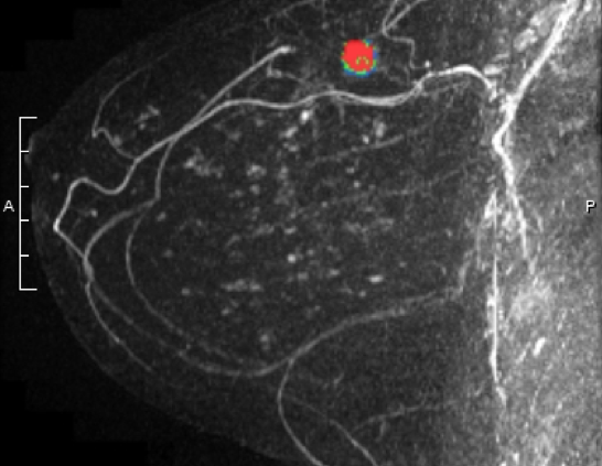
A study recently published in the New England Journal of Medicine* suggests that the use of breast MRI in addition to mammograms in women with extremely dense breast can improve the rate of “interval cancer” detection.

Interval cancers are those that occur after an apparently normal mammogram, but prior to the next scheduled mammogram. An example could be a lump felt by a patient a few months after a “clear” mammogram. Interval cancers are more likely to be more aggressive (triple negative or Her2/neu over-expressed) and/or occur in women who have dense breasts on mammogram.

Researchers in the Netherlands evaluated the role of breast MRI as a supplemental screening tool in women age 50-74, who had a normal mammogram and an “extremely dense” (BIRADS 4 density) reading. After a normal mammogram, women were randomized 1:4 to supplemental MRI versus continued mammogram only (1 woman received an invitation for MRI for every 4 who continued undergoing only mammograms). The standard mammography screening in the Netherlands is every 2 years. 8061 women were in the “intent to treat” with MRI group and 32,312 in the mammogram alone group. 59% (4783) of women randomized to the MRI group actually accepted and underwent supplemental MRI.

Some of the key findings:

  • In the MRI “intent to treat” group, they found 2.5 / 1000 interval cancers and in the mammography group there were 5.0 / 1000 interval cancers
  • However, out of the 20 interval cancers discovered in the MRI “intent to treat” group, only 4 were in women who had actually undergone MRI. The other 16 were in women were randomized to the MRI group but did not have the study – they had mammograms only
  • Out of the women who underwent MRI, approximately 10% (454) were recalled for additional imaging
  • 300 women had needle biopsies due to findings on MRI, and out of those, 79 (26%) were found to have cancer. 64 of the cancers were invasive and 15 were noninvasive (DCIS). 74% of the biopsies did NOT show cancer
  • The cancers detected by MRI on average were smaller than those detected by mammography. The majority were hormone receptor positive and lymph node negative
  • DCIS was more likely to be detected in women undergoing MRI (0.31%) versus mammogram alone (0.03%)
  • 0.1% of patients who had an MRI experienced an adverse reaction including fainting, contrast (dye) reaction, and issues with the intravenous line
  • The study was NOT designed to determine if MRI improved the survival rates from the cancers detected.

Some important points were raised in the accompanying editorial by Dr. Dan Longo*. He noted that with any screening test, the goal should be that early detection of the cancer results in a higher likelihood of cure but admitted that any screening test is associated with false positives, and many patients with an abnormal test will not have cancer. He also noted that an “imprecisely defined fraction of cancers that are detected by screening do not progress to cause symptomatic disease or to kill the patient” but that “our dilemma is that for most tumors we cannot tell the difference between cancers that can kill you and those that cannot.”

The study focused on patients with extremely dense breasts, which the authors estimate account for approximately 10% of patients undergoing mammograms. It is not clear if these findings would be similar in patients with level 3 breast density (heterogeneously dense), those undergoing tomosynthesis (3D) mammograms, or in patients who are undergoing mammograms every year. They did not stratify patients by risk assessment and it’s important to note that while dense breasts are a risk factor for breast cancer, not all women with dense breasts are considered “high risk”. US national guidelines currently recommend supplemental imaging with MRI in patients who have a lifetime risk of breast cancer of 20% or greater as calculated by a risk assessment model. Due to high recall and false positive rates associated with MRI as well as possible concerns related to the contrast material, we do not currently recommend MRI for all women, or even for all women with dense breasts.

As one of their concluding statements, the authors did note that the 10% recall rate “is a concern for potential implementation of supplemental screening” [with MRI]. This study adds to the body of literature regarding cancer detection in women with dense breasts, but Dr. Longo concluded in his editorial: “… will we be putting these women at increased risk of procedures without contributing to their eventual survival?” Going forward, it will be important to look at ways to decrease false positive results and costs of supplemental imaging, as well as evaluate whether or not outcomes are improved by additional testing.

*If you are not able to access the full study and would like a copy, please email me: contact at drattai dot com

21 October 2019

Especially during October, when everything seems to be painted pink, it’s easy to overlook the fact that breast cancer is a disease of women and men. Male breast cancer accounts for 0.6 – 1.0% of all breast cancer cases. In the US, approximately 2600 men will be diagnosed with breast cancer each year. The lifetime risk is about 1 in 1000, versus 1 in 8 for women. Male breast cancer accounts for approximately 500 deaths in the US per year. Risk factors include increasing age, family history including BRCA gene mutations, obesity, alcohol intake, prior chest wall radiation, and low androgen hormone levels.

Male breast cancer tends to be diagnosed in later stages compared with breast cancer in women, and previous studies have come to conflicting conclusions about whether the poorer outcomes are due to higher stage at diagnosis or other factors. A study recently published in JAMA Oncology* looked at mortality rates among men and women diagnosed with breast cancer. The researchers used the National Cancer Database (NCDB) and compared men and women who were diagnosed with breast cancer between January 2004 – December 2014. Their data analysis included approximately 16,000 men and 1.8 million women. Some of the key findings:

  • Mean age at diagnosis was 63.3 for men and 59.9 for women
  • 3-year survival was 86.4% for men and 91.7% for women
  • 5-year survival was 77.6% for men and 86.4% for women
  • Overall survival was 45.8% for men and 60.4% for women

Men diagnosed with breast cancer were older, were more likely to be diagnosed at advanced stages, and were less likely to receive conventional therapy. However, differences in survival persisted even after controlling for clinical characteristics of the disease, age, race and ethnicity, and access to care. Limitations of this study are that cause of death could not be determined (so it is not clear if all of the deaths are related to breast cancer) and the NCDB does not contain information on recurrence, BRCA gene status, adherence to treatment recommendations, and other medical conditions. However, the researchers concluded that male sex remained a significant risk factor for poorer outcomes, which suggests that there are biological differences in male versus female breast cancer. 

Another study recently published in the journal Cancer* also used NCDB information to look at treatment trends for men treated for breast cancer from a similar time period. The authors evaluated approximately 10,000 cases and noted that:

  • 24% underwent breast conserving surgery (lumpectomy)
  • 70% of those undergoing lumpectomy received radiation
  • 44% of patients received chemotherapy
  • 62% of those with estrogen receptor positive (ER+) breast cancer received endocrine therapy
  • 35% of those with ER+ / lymph node negative breast cancer had Oncotype Dx testing on their tumor to help determine need for chemotherapy

These findings are consistent with a point made in the JAMA Oncology study noting that men were less likely to receive conventional therapy – for example only 62% with ER+ breast cancer received endocrine therapy and only 70% of those undergoing breast conserving surgery were treated with postoperative radiation therapy. Some of the same limitations apply to this study, in that reasons for differences in therapy could not be determined, and there was no information on disease recurrence.

A few other important points to make about male breast cancer:

  • Most male breast cancer presents as a lump, but as in women, most lumps are not cancerous. It is important that a proper evaluation (usually including a mammogram and ultrasound, and possibly biopsy) be performed for any change
  • As in women, male breast cancer may present with nipple discharge (especially blood), “puckering” or “pulling in” of the skin, or severe redness of the skin which can be mistaken for infection – the latter may indicate a more aggressive type of breast cancer known as inflammatory breast cancer
  • ALL men with breast cancer, and anyone with a family history of male breast cancer, should undergo genetic counseling and testing. As in women, most cases of male breast cancer are “sporadic” (not related to an inherited mutation), but men with breast cancer are more likely to carry deleterious BRCA (especially BRCA 2) mutations
  • Men who carry a deleterious BRCA mutation have an approximately 8% lifetime risk (to age 80) of developing breast cancer. So while that is considered “high risk” for men, they are still more likely to NOT develop breast cancer. We do not currently recommend prophylactic mastectomy in men who carry a deleterious BRCA mutation but who have not been diagnosed with breast cancer
  • Men who carry a deleterious BRCA mutation are also at higher risk for prostate cancer, melanoma, and pancreatic cancer

Men with breast cancer are usually treated using the same protocols that are used for women. Unfortunately there is limited data to support this. Male breast cancer is not common, so it is challenging to enroll large numbers of patients in clinical trials. However, men have historically been excluded from many breast cancer clinical trials, so how can we even make progress? The US FDA has recently issued draft guidelines encouraging the inclusion of male breast cancer patients in clinical trials – this is certainly a step in the right direction.

*If you are not able to access the full study and would like a copy, please email me: contact at drattai dot com

Additional Information:

14 July 2019

A study recently published in the journal Cancer* reports on disparities in triple negative breast cancer (TNBC). The researchers used the US cancer statistics database, and compared differences between TNBC and other breast cancer subtypes focusing on age, race / ethnicity, and stage at diagnosis. 

Between 2010 and 2014, approximately 1.15 million breast cancer cases were identified in the database. The mean age at diagnosis was 61.8, and 75% of patients were non-Hispanic white (NHW). 27.7% were diagnosed at stage III and 5% were diagnosed at stage IV. 

TNBC accounted for 8.4% of all breast cancer cases, but accounted for 21.4% of cases in non-Hispanic Black (NHB) women. Women diagnosed with TNBC had a lower mean age at diagnosis versus non-TNBC (59.3 versus 62.1). NHB women accounted for 11.8% of the study population, but 15.3% of TNBC cases. Women diagnosed with TNBC were also more likely to be younger than age 40 and diagnosed at Stage III or IV compared to those with other cancer subtypes. After controlling for late-stage diagnosis and age, NHB women had approximately twice the likelihood of diagnosis with TNBC compared with NHW women.

The researchers noted that their study validated previous literature, and had the advantage of being geographically very broad. Not discussed in this paper is the relationship of TNBC with genetic mutations, specifically mutations in the BRCA1 gene. Among patients with breast cancer, those who carry deleterious BRCA1 mutations are more likely to have TNBC versus BRCA2 carriers or non-carriers, and a diagnosis of TNBC should prompt genetic testing.

This study and others like it point to the need for more research on TNBC, and on breast cancer in black women. Dr. Lisa Newman, a breast surgical oncologist at Cornell, has published extensively on this topic. In this brief video, she notes that socioeconomic factors account for some of the disparities associated with breast cancer outcomes in black women. However, she also notes that there appears to be biologic differences between TNBC in black versus white women, which may also at least in part account for later stage at diagnosis and poorer prognosis.

*If you are not able to access the full study and would like a copy, please email me: contact at drattai dot com

8 July 2019

Patients who have been diagnosed with atypical ductal hyperplasia (ADH) and lobular carcinoma in-situ (LCIS) have a much higher risk of developing breast cancer compared with the average population, usually well over the “high risk” threshold of 20%. Tamoxifen is recommended for these patients to reduce help reduce subsequent breast cancer risk, but a relatively low percentage of patients actually take it, often due to concerns about side effects. 

In December 2018, the TAM-01 study was presented at the San Antonio Breast Cancer Symposium. This study used a reduced dose (5 mg instead of the usual 20 mg daily) of tamoxifen in women who had ADH, LCIS or ductal carcinoma in situ (DCIS) to see if a lower than standard dose could effectively reduce breast cancer risk.

The full manuscript* has recently been published. 500 women with a history of ADH, LCIS or DCIS were randomized to receive 3 years of either low dose tamoxifen or placebo. The study was performed in Italy. Mean patient age was 54, and 55% of the patients were post-menopausal. 20% had ADH, 11% had LCIS, and 69% had DCIS. 

With a median follow up of 5.1 years, there were 14 cancers (invasive or DCIS) in the tamoxifen group and 28 in the placebo group (11.6 versus 23.9 per 1000 person-years). Tamoxifen decreased the number of contralateral (opposite side) events by 75% but numbers were low (3 versus 12). Daily hot flashes in patients receiving tamoxifen were more frequent (2 versus 1.5 per day) than in patients receiving placebo. There were no differences in patient-reported hot flash score (combination of hot flash frequency and intensity), vaginal dryness, pain during sexual intercourse, or joint / muscle pains between the 2 groups. There were fewer serious adverse events (blood clot, pulmonary embolus, uterine cancer) in the patients receiving tamoxifen versus placebo (12 versus 16). 65% of patients in the tamoxifen arm and 61% in the placebo arm took ≥85% of pills for the first 2 ½ years of the study. The authors concluded that low dose tamoxifen for 3 years can reduce the risk of invasive or non-invasive breast cancer, with similar side effects to placebo. 

It is important to stress that this is one study, with a relatively small number of patients – 195 of the 253 in the tamoxifen arm completed the study. An important finding is that the authors found risk-reducing effects after only 3 years of therapy. However, current guidelines do still recommend standard (20mg) tamoxifen for 5 years for high risk patients as well as those with a history of estrogen-receptor positive (ER+) DCIS. There is no data on the use of low dose tamoxifen for invasive breast cancer treatment. However, for patients who are reluctant to take the full dose of tamoxifen, or who have significant side effects, it may be reasonable to consider a reduced dose – taking into account the patient’s individual risk, side effects of the standard 20mg dose, and the limitations of this study.

*Journal of Clinical Oncology Editorial: Will a low-dose option improve uptake of tamoxifen for breast cancer risk reduction?

*If you are not able to access the full study and would like a copy, please email me: contact at drattai dot com

5 July 2019

A study recently published in The Lancet Oncology found that the addition of MRI to mammography screening in high-risk patients detected breast cancers in earlier stages, but found that this technology was associated with a higher likelihood of false-positive results.

The study was performed at 12 hospitals in the Netherlands. Patients who were age 30-55 with a lifetime risk of breast cancer greater than 20% due to family history, but who tested negative for deleterious mutations in BRCA 1/2 and TP53 genes, were eligible to participate. Patients were randomized to one of the following surveillance arms:

  • Annual MRI, annual clinical breast exam, every other year mammogram [MRI-MG], or
  • Annual mammogram and annual clinical breast exam [MG]

1355 women were randomized and 231 were registered – the patients who were registered were those who did not provide consent for randomization, but followed one or the other protocol. Results from the patients who were registered but not randomized were used only in a portion of the data analysis.  

The authors found that over the 7-year study period:

  • 40 breast cancers were detected in the MRI-MG group, 15 were detected in the MG group
  • 24 invasive breast cancers were detected in the MRI-MG group, 8 were detected in the MG group
  • Invasive breast cancers detected in the MRI-MG group were smaller (9 versus 17 millimeters) than those in the MG group
  • Lymph node involvement was less likely to be seen in the MRI-MG group (4 of 24 cases, 17%) versus the MG group (5 of 8 cases, 63%)
  • There was one interval cancer (detected between screening exams) in the MRI-MG group and 2 in the MG group
  • Increased breast density was associated with higher stage at diagnosis in both study groups
  • Clinical breast exams generated a large number of false-positive results in both groups and detected only one cancer.

This study adds to the evidence nothing that the addition of MRI to screening mammography will increase the rate of cancer detection. National guidelines in the US do recommend breast MRI on an annual basis, in addition to annual mammography, for patients with a lifetime risk of 20% or greater. This study was not designed to assess outcomes for the patients who were diagnosed with breast cancer, but the authors postulated that MRI screening might lead to mortality reduction due to earlier stage at diagnosis. As reduction in mortality is the goal of screening, they stated that they plan to link the current study results with their national cancer database, and eventually publish 10-year mortality statistics for each of the groups. 

Additional Information: ASCO Post

3 May 2019

The American Society of Breast Surgeons (ASBrS) has updated their screening mammography guideline, recommending that women of average risk begin annual screening mammograms at age 40. This update brings the ASBrS guideline more in line with those of the American College of Radiology and the Society of Breast Imaging, and are a departure from their previous guideline, which called for screening to start at age 45. The American Cancer Society recommends annual screening from age 45-54, followed by biennial screening with the opportunity for annual screening. The US Preventative Services Task Force recommends biennial screening starting at age 50.

The updated ASBrS guidelines recommend as a first step that all women undergo a formal risk assessment starting at age 25. Risk assessment involves using any one of a number of models to estimate 5-year and lifetime risk of breast cancer development. These models take into account age, family history, menstrual and pregnancy history as well as history of prior biopsy and racial / ethnic background. Some models take into account body mass index and breast density. The ASBrS guideline has additional recommendations, outlined below, depending on risk level. The recommendations listed for women at high risk of breast cancer are consistent with those of the American Cancer Society and National Comprehensive Cancer Network.

When to start and how often to perform screening mammography has been the subject of much debate over the years. The primary issue is the number of lives saved balanced against the harms of screening mammography. Harms include false negatives (cancer is not detected by the mammogram), false positives (suspicious areas that turn out to be benign) and recommendations for additional imaging (added cost and concerns about the potential effects of cumulative radiation). There have also been concerns raised about over-diagnosis and over-treatment: finding cancers that would never pose a threat to a woman’s health or life – but the patient is exposed to the potential harms of cancer therapy.

The goal of screening is not to detect all cancers – otherwise we would recommend complete body imaging for everyone. The goal of screening is to improve the survival from the cancers that are detected. An ideal screening test is relatively inexpensive and performs equally well in the patient population undergoing the screening. Mammography is acknowledged to be an imperfect screening tool – while relatively inexpensive and safe, it simply does not perform the same in women with breasts of different ages and densities. Approximately 1000 women need to be screened to detect 2 – 7 breast cancers, and mammography performs best in women age 50-74. In younger women, a few cancers will be detected, and there may be a survival benefit. However, because breast cancer is less common in younger women, a larger number of women need to be screened to find a single cancer, increasing the likelihood of a harm. The benefits of screening do not always outweigh the risks – at least for an individual patient.

It is also important to note that some of the new ASBrS recommendations, such as 3D mammography as the preferred type of mammogram, supplemental ultrasound in women with dense breasts, and MRI for all women with a history of breast cancer, are still undergoing investigation and are not always covered by insurance. We know that these studies will find additional cancers, but we do not yet have data on improved outcomes. My patients know that I am fond of saying “the more we look, the more we find, but not everything we find needs to be found.”

Two ongoing studies will hopefully provide additional information regarding 3D mammography and screening interval:

  • The TMIST trial is a national multi-center study assessing the performance of 3D compared to 2D digital mammography. This study will assess rates of cancer detection, callbacks and benign biopsies as well as biology of the tumors detected and outcomes.
  • The WISDOM study is evaluating a risk-based screening approach. After a comprehensive risk assessment, genetic testing, and assessment of risk tolerance, patients are assigned annual versus biennial screening. Women can self-enroll in this study (disclosure – I am not involved with the study team but I am enrolled in the study as a participant) and do not have to change mammography facility.

Until we are at a point when an individual woman’s level of risk can be accurately predicted, there is no definitive “best” screening guideline to follow. Recognizing the potential harms of screening mammography, it is no longer appropriate for physicians to simply hand out an order for an annual screening mammogram for all women starting at age 40. A balanced discussion, taking into account an individual woman’s risk and level of risk tolerance as well as the absolute potential benefits and harms of mammography is indicated. These discussions are nuanced and take time. Various genomic assays are being evaluated and it is hopeful that we will at some point have a test that can accurately predict a woman’s risk of breast cancer – which can then be used to provide more tailored guidance regarding imaging. Until that time (and I hate to end this way…), talk to your physician and medical team about what screening option is right for you.

1 January 2018

The American Joint Committee on Cancer (AJCC) has recently updated the staging system used for breast cancer. Cancer stage refers to the amount of cancer (size of the main tumor, spread to lymph nodes or other areas). The definitions of each stage vary depending on cancer type. Cancer stage often correlates with outcomes, and treatment recommendations usually take into account stage of disease.

As our knowledge of tumor biology increases, it has become clear that stage is not the only factor that impacts prognosis. Tumor biology and behavior are very important, and in some cases may be more important than stage. A small tumor with aggressive behavior characteristics may may result in worse outcomes compared to a larger, but slower growing cancer. The 8th version of the AJCC staging system for breast cancer now takes into account tumor biology. Factors such as cell grade, ER/PR and Her2/neu status, and even the results of tumor genomic tests will be incorporated into the clinical and pathological prognostic stage. Taking into account these biologic factors means that the stage will have more meaningful prognostic information. Some larger tumors will now be considered stage I, and some smaller lesions will be upstaged based on their biology. In a large validation study performed by researchers at the University of Texas MD Anderson Cancer Center, 31% of patients were upstaged, and 20% of patients were downstaged. The updated prognostic stage performed better (in terms of predicting patient outcomes) than the standard anatomic stage.

The new staging system will take some getting used to. The tables used to help determine clinical and pathologic prognostic stage are 5-6 pages long. However, this new system will give us more meaningful information in terms of prognosis and outcomes. Just please be patient with your physician when you ask “what is my stage?” – it is no longer a simple question!

Additional Information:
American Cancer Society CA Cancer Journal: Breast Cancer – Major Changes in the AJCC 8th Edition

21 June 2016

In October, we solicited input a research survey designed to identify how patients prefer to be notified of a new cancer diagnosis and cancer-related test results. The results were presented at the 2016 Annual Meeting of the American Society of Breast Surgeons in April. The full manuscript – What Do Patients Prefer? Understanding Patient Preferences for Receiving a New Breast Cancer Diagnosis – was just published in the Annals of Surgical Oncology.

Our study showed that physicians are not meeting patient expectations regarding mode of diagnosis disclosure (phone versus in person) or timeliness of results disclosure. The majority of patients who responded to our survey preferred to be notified in person, however many commented that they truly preferred the method that was fastest. More patients preferred to be notified of radiology results by telephone, and electronic methods such as email or patient portal were preferred for blood test results.

We included many of your comments in the manuscript, and it is clear that one size most definitely does not fit all in terms of results disclosure. However, from our study it is very clear that we are not meeting patient expectations. System issues should be evaluated to identify areas of improvement in terms of timeliness of results disclosure, and physicians should ask patients how they prefer to be notified, and should make every effort to honor patient preferences.

Thank you to the community for your participation in this study, and for helping to make an important contribution to the literature. This study would not have happened without the support of the #BCSM community, as well as the Dr. Susan Love Research Foundation, Living Beyond Breast Cancer, and the Seattle Cancer Care Alliance. If anyone is interested in receiving the manuscript (and you are not able to download it from the Springer website) please email me: [contact at drattai dot com] and I will be happy to send you a copy of the paper.
Deanna J. Attai MD, FACS
Regina Hampton MD, FACS
Alicia Staley BS, MBA, MS
Andrew Bogert PhD
Jeffrey Landercasper MD, FACS