10 May 2018
The American Society of Breast Surgeons held their Annual Meeting in Orlando, FL from May 2nd – 6th. As usual, it was well attended – the meeting is known for being very practical and full of information that breast surgeons can bring back to their practices to help improve patient care.
I’ve picked a few topics to highlight in this post: Genetics, Imaging, Local Therapy, Systemic Therapy, Immunotherapy, Liquid Biopsy, Diet and Hormone Therapy, and Changing Paradigms. The following are comments expressed by the meeting speakers. My own comments will be noted in bold italics.
Genetics:
- BRCA 1 mutation carriers are more likely to have triple negative breast cancer.
- BRCA 2 mutation carriers are more likely to have ER positive, Her2/neu negative breast cancers.
- The risk of a 2nd breast cancer in BRCA mutation carriers on average is about 2% per year depending on the specific mutation and the age of affected relatives. It can approach 60-80% in some patients. This increased risk of a new breast cancer is why bilateral mastectomy is often recommended. Removal of the opposite breast may result in improved overall survival but results from studies are mixed.
- For BRCA mutation carriers, it is recommended that clinical breast exam (breast exam by the physician) be performed every 6-12 months. From age 25-29 annual MRI is recommended, and from age 30-75 annual mammogram (3D mammogram or tomosynthesis was recommended) along with MRI was recommended. It was stated that this screening regimen has not been shown to improve survival, but the screen-detected cancers were less likely to have lymph node involvement. No specific recommendation was made for imaging or exam after bilateral mastectomy.
- MRI every 6 months has been suggested by some, but there are concerns about gadolinium (a heavy metal material which is the contrast agent used for breast MRI) buildup.
- Removal of the ovaries is recommended around age 40.
- In patients with BRCA mutations who undergo salpingo-oophorectomy (removal of the ovaries and fallopian tubes), estrogen replacement therapy has not been shown to increase subsequent breast cancer risk. However, combined estrogen / progesterone therapy may increase subsequent breast cancer risk. It was suggested to consider removing the uterus at the time of ovary removal, so that estrogen alone could be used (if the uterus is not removed, estrogen alone could increase the risk of uterine cancer).
- There are many other genetic mutations that have been identified that have a variable association with increased breast cancer risk. It was stressed that family history and other factors need to be considered when these less common mutations (such as CHEK2, ATM, PALB2 and many more) are present, before recommending mastectomy.
- It was stressed that the presence of a variant of unknown significance (VUS) should NOT prompt aggressive surgery.
- A study was presented that demonstrated that current breast cancer genetic testing guidelines exclude almost half of high-risk patients, and a recommendation was made for testing of all breast cancer patients regardless of age, family history or other factors.
Breast Imaging:
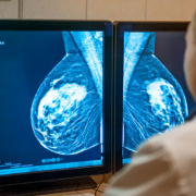
- Dense breast (as determined by mammogram) reduces the sensitivity of mammograms, and also is associated with an increased risk of breast cancer.
- It was stressed that determination of breast density is subjective and studies have shown significant variability in grading of breast density. Automated methods of assessing density are being evaluated.
- 34 states have dense breast notification legislation. Some have supplemental screening (such as ultrasound) legislation (California does not).
- An advantage of tomosynthesis (also known as 3D mammogram) in patients with dense breasts is that it decreases the likelihood of callbacks and improves the cancer detection rate
- Abbreviated (3 minute scan) MRI shows promise for screening.
- There is an ECOG/ACRIN study planned which will evaluate abbreviated MRI versus tomosynthesis in women with dense breasts.
- Contrast-enhanced mammography is superior to digital mammography but it requires an IV contrast dye, and there is currently no ability to biopsy lesions seen only with this technique.
- It was stressed that automated whole breast ultrasound (ABUS) should not replace mammography.
- Molecular breast imaging has a much higher radiation dose due to the need to inject a radioactive material and cost is higher than other imaging modalities. There are only about 100 units in the US.
- In addition to BRCA mutation carriers, patients who have a history of chest wall radiation at a young age (most commonly for treatment of Hodgkin’s lymphoma) or those who have a lifetime risk of breast cancer over 20% (assessed by various computer modes) should have annual MRI in addition to mammograms for surveillance.
Loco-Regional (breast and underarm lymph nodes) Therapy:
- Recurrence of cancer in the breast (known as a local recurrence) was previously thought to be related to “disease burden” – the amount of tumor and size of clear margins. According to Dr. Monica Morrow, this has led to an “obsession” with margins, wider surgical resection than necessary, and the overuse of MRI.
- Due to improvements in systemic therapy (chemotherapy and endocrine therapy), local recurrences have decreased over time.
- Local recurrences are largely a function of tumor biology – more aggressive tumor types are more likely to recur. Bigger surgery does not overcome bad biology.
- The rates of contralateral (opposite side) new breast cancer have been decreasing in the US; currently <1% at 5 years for patients who do not have a genetic mutation.
- Updated 2018 ASTRO guidelines endorse hypofractionation (a shorter course of radiation therapy) in a larger group of patients.
- There are 3 trials that will evaluate whether or not radiation therapy can be avoided in selected patients – LUMINA, IDEA and PRECISION.
- ~30% of patients undergoing “direct to implant” reconstruction (no temporary tissue expander) need a second surgery. One of the plastic surgeons that I work with notes that “reconstruction is a process not a procedure!”
- Managing expectations of the reconstruction process is important so patients don’t get frustrated and feel like their reconstruction has “failed.”
- Post mastectomy radiation worsens outcome from implant reconstruction; severe capsular contracture occurs in about 30% of patients.
- If radiation is performed on the permanent implant instead of the tissue expander, the rate of reconstruction failure goes down by 50%.
- Many plastic surgeons prefer that autologous (patient’s own body) reconstruction be performed after radiation to avoid shrinkage of the flap. A tissue expander could be placed at the time of mastectomy which will be removed after radiation when the flap procedure is performed.
- Lymphedema risk is about 25% with axillary node dissection versus 6-8% with sentinel node biopsy. In certain patients over age 70 with ER+ breast cancer, sentinel node biopsy can be avoided – this was also covered in the Society of Surgical Oncology’s Choosing Wisely statements. However, it is also important to take into account whether or not the patient will be treated with radiation and/or endocrine therapy. Sentinel node biopsy is also not recommended for most patients undergoing lumpectomy for DCIS. The SOUND trial is evaluating the use of axillary ultrasound to try to determine if this can help select patients who do not need sentinel node biopsy.
Systemic Therapy:
- The use of genomic tumor testing could avoid the use of ineffective (for the specific patient depending on tumor profile) chemotherapy in up to 50,000 patients per year.
- Neoadjuvant (before surgery) chemotherapy is most commonly used to decrease tumor size so that patients have a higher likelihood of being able to undergo lumpectomy instead of mastectomy.
- About 50% of patients who have positive lymph nodes before chemotherapy are converted to node-negative due to chemotherapy prior to surgery, and they may be able to avoid full axillary node dissection.
- Response to neoadjuvant chemotherapy varies by tumor subtype. Her2/neu and triple negative breast cancers are more likely to respond compared to ER+ and Her2/neu negative tumors.
- Technical considerations to improve the accuracy of sentinel node biopsy after neoadjuvant chemotherapy including the use of 2 dye agents to map the nodes and removal of at least 3 lymph nodes.
- A multidisciplinary approach for management of patients who are being considered for neoadjuvant chemotherapy was stressed.
- Recurrence patterns are different for ER+ versus ER- disease. Patients with ER+ breast cancer are at risk for late recurrence, even 20 years after treatment – the highest risk is in patients with multiple involved lymph nodes. Patients with ER- disease tend to recur earlier (within the first 2-5 years), and then the likelihood of recurrence decreases.
- Recurrence in the breast is a marker of increased risk for development of metastatic disease.
- Premenopausal patients who have “low risk” disease could consider stopping tamoxifen after 5 years. It is recommended that patients with “high risk” disease consider 10 years of tamoxifen therapy.
- Postmenopausal patients who are considered “high risk” could consider 10 years of an aromatase inhibitor, although there is not currently data that shows this approach improves survival. Prolonged therapy in these patients does reduce the likelihood of developing a new breast cancer and reduces the likelihood of breast cancer recurrence.
Immunotherapy / Liquid Biopsy:
- A brief session was held covering immunotherapy and liquid biopsy.
- Immunotherapy for breast cancer has not had the success seen in melanoma, lung cancer, colon cancer and bladder cancer.
- The combination of chemotherapy and a modified herpes virus has shown some promise in patients with triple negative breast cancer.
- It is likely that immunotherapy treatments will vary depending on tumor subtype.
- Circulating tumor DNA may predict metastatic disease 8-12 months before evidence of tumor spread – but we are not yet able to improve patient outcomes based on this information. Therefore, circulating tumor cell and circulating cancer cell DNA assessments are not recommended for routine clinical use.
- It was predicted that “liquid biopsy” will eventually be used routinely to help manage breast cancer patients.
Diet and Hormone Replacement Therapy:
- A low fat diet improved the likelihood of death from breast cancer only in obese women.
- Currently there is more information regarding the impact of dietary fat versus dietary sugar on breast cancer risk. Dr. Rowan Chlebowski, who has been a lead author on the Women’s Health Initiative studies, stated that due to an increasing number of reports suggesting that sugar may impact breast cancer development, they plan to look more closely at this.
- Insulin resistance is associated with cancer specific and all-cause mortality in postmenopausal women.
- One of Dr. Chlebowski’s conclusions was to “avoid body fatness.” Unfortunately, specific guidance on how to best accomplish this was not discussed!
- The risk of breast cancer associated with hormone replacement therapy (HRT) is greater if it is started around the time of menopause versus 3-5 years later.
- Breast cancer risk in women taking HRT is higher in women with extremely dense breast versus fatty replaced breasts. The biggest risk from HRT is in lean women with extremely dense breasts. The lowest risk from HRT is in women with a body mass index (BMI) > 35 with fatty replaced breasts.
- Combination estrogen / progesterone HRT should be avoided in lean (BMI <25) women especially if they have dense breast tissue.
- The Black Women’s Health Study found no increased breast cancer risk if HRT use was <10 years, but cancer risk was increased if use was >10 years. Other studies showed either no risk or no association of risk from HRT with race.
Changing Paradigms – Avoiding Surgery for DCIS and Neoadjuvant Patients
- Active surveillance is being evaluated for ductal carcinoma in-situ (DCIS). Over 60,000 cases of DCIS are diagnosed per year in the US. Not all cases of DCIS will progress to invasive cancer, and the likelihood of progression is lowest in low grade DCIS. In these patients, less than 10% develop invasive cancer in the same breast after 10 years and over 20% die from other causes within 10 years of diagnosis.
- There are 3 ongoing clinical trials are evaluating active surveillance for low risk DCIS (LORIS, LORD, and COMET). The COMET trial is the only study open in the US. DCISOptions.org has additional information about DCIS and the COMET trial.
- Some patients who undergo chemotherapy prior to surgery are found to have no residual tumor after the area has been removed, termed pathologic complete response (pCR).
- Prompted by patients asking “why do I need surgery?” if it appears that all cancer has resolved after chemotherapy, researchers at MD Anderson Cancer Center are evaluating whether surgery can be omitted in patients who appear to have a pCR after chemotherapy. Patients who have no apparent tumor based on post-chemotherapy imaging (including MRI) undergo core needle biopsies. If these biopsies show no tumor, patients taking part in the study will undergo radiation without surgery.
- Similar studies are taking place in the Netherlands, Germany, and the UK.
- Henry Kuerer from MD Anderson stated that “surgeons have an obligation to study possibility of no surgery – and we must ensure safety and efficacy with well-designed trials.
- Several types of ablative therapy (destroying the tumor without surgery) are being evaluated including cryoablation (freezing), laser, and transcutaneous (no needle puncture or scar) high frequency ultrasound.
Lifetime Achievement Award
Dr. Ernie Bodai, the breast surgeon who spearheaded the Breast Cancer Research Stamp, was honored with a lifetime achievement award. It was fascinating to hear his story and how one man (with a little help) got congress to change a law.
This post has not been endorsed by the American Society of Breast Surgeons


